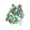
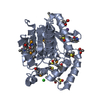
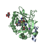
[English] 日本語
Yorodumi
- PDB-6t7h: Crystal structure of Thrombin in complex with macrocycle N14-PR4-A -

+
Open data


ID or keywords:

Loading...

-
Basic information

Entry
Database: PDB / ID: 6t7h
TitleCrystal structure of Thrombin in complex with macrocycle N14-PR4-A
Components
  • Thrombin heavy chain
  • Thrombin light chain
KeywordsHYDROLASE / serine protease / blood clotting factor / inhibition / macrocycle
Function / homology
Function and homology information


positive regulation of lipid kinase activity / positive regulation of phospholipase C-activating G protein-coupled receptor signaling pathway / cytolysis by host of symbiont cells / thrombospondin receptor activity / Defective factor XII causes hereditary angioedema / thrombin / neutrophil-mediated killing of gram-negative bacterium / regulation of blood coagulation / ligand-gated ion channel signaling pathway / Defective F8 cleavage by thrombin ...positive regulation of lipid kinase activity / positive regulation of phospholipase C-activating G protein-coupled receptor signaling pathway / cytolysis by host of symbiont cells / thrombospondin receptor activity / Defective factor XII causes hereditary angioedema / thrombin / neutrophil-mediated killing of gram-negative bacterium / regulation of blood coagulation / ligand-gated ion channel signaling pathway / Defective F8 cleavage by thrombin / Platelet Aggregation (Plug Formation) / negative regulation of platelet activation / negative regulation of astrocyte differentiation / negative regulation of cytokine production involved in inflammatory response / positive regulation of collagen biosynthetic process / positive regulation of blood coagulation / negative regulation of fibrinolysis / Gamma-carboxylation of protein precursors / Transport of gamma-carboxylated protein precursors from the endoplasmic reticulum to the Golgi apparatus / Common Pathway of Fibrin Clot Formation / Removal of aminoterminal propeptides from gamma-carboxylated proteins / regulation of cytosolic calcium ion concentration / fibrinolysis / Intrinsic Pathway of Fibrin Clot Formation / Peptide ligand-binding receptors / positive regulation of release of sequestered calcium ion into cytosol / Regulation of Complement cascade / acute-phase response / Cell surface interactions at the vascular wall / lipopolysaccharide binding / negative regulation of proteolysis / positive regulation of receptor signaling pathway via JAK-STAT / growth factor activity / positive regulation of insulin secretion / platelet activation / response to wounding / Golgi lumen / positive regulation of protein localization to nucleus / Regulation of Insulin-like Growth Factor (IGF) transport and uptake by Insulin-like Growth Factor Binding Proteins (IGFBPs) / positive regulation of reactive oxygen species metabolic process / blood coagulation / antimicrobial humoral immune response mediated by antimicrobial peptide / Thrombin signalling through proteinase activated receptors (PARs) / heparin binding / regulation of cell shape / positive regulation of cell growth / G alpha (q) signalling events / collagen-containing extracellular matrix / blood microparticle / cell surface receptor signaling pathway / positive regulation of phosphatidylinositol 3-kinase/protein kinase B signal transduction / positive regulation of protein phosphorylation / G protein-coupled receptor signaling pathway / endoplasmic reticulum lumen / signaling receptor binding / serine-type endopeptidase activity / calcium ion binding / positive regulation of cell population proliferation / proteolysis / extracellular space / extracellular exosome / extracellular region / plasma membrane
Similarity search - Function
Prothrombin/thrombin / Thrombin light chain / Thrombin light chain domain superfamily / Thrombin light chain / Kringle domain / Kringle / Kringle, conserved site / Kringle superfamily / Kringle domain signature. / Kringle domain profile. ...Prothrombin/thrombin / Thrombin light chain / Thrombin light chain domain superfamily / Thrombin light chain / Kringle domain / Kringle / Kringle, conserved site / Kringle superfamily / Kringle domain signature. / Kringle domain profile. / Kringle domain / Vitamin K-dependent carboxylation/gamma-carboxyglutamic (GLA) domain / Gamma-carboxyglutamic acid-rich (GLA) domain / Gamma-carboxyglutamic acid-rich (GLA) domain superfamily / Vitamin K-dependent carboxylation domain. / Gla domain profile. / Domain containing Gla (gamma-carboxyglutamate) residues. / Kringle-like fold / Serine proteases, trypsin family, histidine active site / Serine proteases, trypsin family, serine active site / Peptidase S1A, chymotrypsin family / Serine proteases, trypsin family, histidine active site. / Serine proteases, trypsin domain profile. / Serine proteases, trypsin family, serine active site. / Trypsin-like serine protease / Serine proteases, trypsin domain / Trypsin / Peptidase S1, PA clan, chymotrypsin-like fold / Peptidase S1, PA clan
Similarity search - Domain/homology
Chem-MRQ / Prothrombin
Similarity search - Component
Biological speciesHomo sapiens (human)
MethodX-RAY DIFFRACTION / SYNCHROTRON / MOLECULAR REPLACEMENT / molecular replacement / Resolution: 2.32 Å
AuthorsAngelini, A. / Kumar, M.G. / Heinis, C. / Cendron, L.
Funding support Switzerland, 1items
OrganizationGrant numberCountry
Swiss National Science Foundation157842 Switzerland
CitationJournal: Chem Sci / Year: 2020
Title: Macrocycle synthesis strategy based on step-wise "adding and reacting" three components enables screening of large combinatorial libraries.
Authors: Mothukuri, G.K. / Kale, S.S. / Stenbratt, C.L. / Zorzi, A. / Vesin, J. / Bortoli Chapalay, J. / Deyle, K. / Turcatti, G. / Cendron, L. / Angelini, A. / Heinis, C.
History
DepositionOct 22, 2019Deposition site: PDBE / Processing site: PDBE
Revision 1.0Sep 30, 2020Provider: repository / Type: Initial release
Revision 1.1Jun 16, 2021Group: Database references / Category: citation / citation_author
Item: _citation.journal_id_ISSN / _citation.journal_volume ..._citation.journal_id_ISSN / _citation.journal_volume / _citation.page_first / _citation.page_last / _citation.pdbx_database_id_PubMed / _citation.title / _citation_author.name
Revision 2.0Sep 8, 2021Group: Advisory / Atomic model ...Advisory / Atomic model / Data collection / Database references / Derived calculations / Polymer sequence / Structure summary
Category: atom_site / chem_comp ...atom_site / chem_comp / database_2 / entity / entity_name_com / entity_poly / pdbx_entity_nonpoly / pdbx_nonpoly_scheme / pdbx_poly_seq_scheme / pdbx_struct_conn_angle / pdbx_struct_sheet_hbond / pdbx_unobs_or_zero_occ_residues / pdbx_validate_torsion / struct_conf / struct_conn / struct_mon_prot_cis / struct_ref_seq / struct_sheet_range
Item: _atom_site.auth_asym_id / _chem_comp.name ..._atom_site.auth_asym_id / _chem_comp.name / _chem_comp.pdbx_synonyms / _chem_comp.type / _database_2.pdbx_DOI / _database_2.pdbx_database_accession / _entity.pdbx_description / _entity_poly.pdbx_strand_id / _pdbx_entity_nonpoly.name / _pdbx_nonpoly_scheme.pdb_strand_id / _pdbx_poly_seq_scheme.pdb_strand_id / _pdbx_struct_conn_angle.ptnr1_auth_asym_id / _pdbx_struct_conn_angle.ptnr2_auth_asym_id / _pdbx_struct_conn_angle.ptnr3_auth_asym_id / _pdbx_struct_sheet_hbond.range_1_auth_asym_id / _pdbx_struct_sheet_hbond.range_2_auth_asym_id / _pdbx_unobs_or_zero_occ_residues.auth_asym_id / _pdbx_validate_torsion.auth_asym_id / _struct_conf.beg_auth_asym_id / _struct_conf.end_auth_asym_id / _struct_conn.ptnr1_auth_asym_id / _struct_conn.ptnr2_auth_asym_id / _struct_mon_prot_cis.auth_asym_id / _struct_mon_prot_cis.pdbx_auth_asym_id_2 / _struct_ref_seq.pdbx_strand_id / _struct_sheet_range.beg_auth_asym_id / _struct_sheet_range.end_auth_asym_id
Revision 2.1Jan 24, 2024Group: Data collection / Refinement description
Category: chem_comp_atom / chem_comp_bond / pdbx_initial_refinement_model

-
Structure visualization

Structure viewerMolecule:
MolmilJmol/JSmol

Downloads & links

-
Assembly

Deposited unit
A: Thrombin light chain
B: Thrombin heavy chain
L: Thrombin light chain
H: Thrombin heavy chain
hetero molecules


Theoretical massNumber of molelcules
Total (without water)69,67413
Polymers67,7544
Non-polymers1,9209
Water2,756153
1
A: Thrombin light chain
B: Thrombin heavy chain
hetero molecules


Theoretical massNumber of molelcules
Total (without water)34,8687
Polymers33,8772
Non-polymers9915
Water362
TypeNameSymmetry operationNumber
identity operation1_555x,y,z1
Buried area2680 Å2
ΔGint-15 kcal/mol
Surface area13270 Å2
MethodPISA
2
L: Thrombin light chain
H: Thrombin heavy chain
hetero molecules


Theoretical massNumber of molelcules
Total (without water)34,8066
Polymers33,8772
Non-polymers9294
Water362
TypeNameSymmetry operationNumber
identity operation1_555x,y,z1
Buried area3150 Å2
ΔGint-18 kcal/mol
Surface area13260 Å2
MethodPISA
Unit cell
Length a, b, c (Å)64.664, 101.343, 119.129
Angle α, β, γ (deg.)90.000, 90.000, 90.000
Int Tables number19
Space group name H-MP212121

-
Components

-
Protein/peptide / Protein / Sugars , 3 types, 6 molecules ALBH

#1: Protein/peptide Thrombin light chain /


Mass: 4096.534 Da / Num. of mol.: 2 / Source method: isolated from a natural source / Details: Missing density / Source: (natural) Homo sapiens (human) / References: UniProt: P00734, thrombin
#2: Protein Thrombin heavy chain /


Mass: 29780.219 Da / Num. of mol.: 2 / Source method: isolated from a natural source / Details: Missing density / Source: (natural) Homo sapiens (human) / References: UniProt: P00734, thrombin
#3: Sugar ChemComp-NAG / 2-acetamido-2-deoxy-beta-D-glucopyranose / N-acetyl-beta-D-glucosamine / 2-acetamido-2-deoxy-beta-D-glucose / 2-acetamido-2-deoxy-D-glucose / 2-acetamido-2-deoxy-glucose / N-ACETYL-D-GLUCOSAMINE / N-Acetylglucosamine


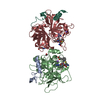
Type: D-saccharide, beta linking / Mass: 221.208 Da / Num. of mol.: 2
Source method: isolated from a genetically manipulated source
Formula: C8H15NO6
IdentifierTypeProgram
DGlcpNAcbCONDENSED IUPAC CARBOHYDRATE SYMBOLGMML 1.0
N-acetyl-b-D-glucopyranosamineCOMMON NAMEGMML 1.0
b-D-GlcpNAcIUPAC CARBOHYDRATE SYMBOLPDB-CARE 1.0
GlcNAcSNFG CARBOHYDRATE SYMBOLGMML 1.0

-
Non-polymers , 4 types, 160 molecules

#4: Chemical ChemComp-EDO / 1,2-ETHANEDIOL / ETHYLENE GLYCOL / Ethylene glycol


Mass: 62.068 Da / Num. of mol.: 3 / Source method: obtained synthetically / Formula: C2H6O2
#5: Chemical ChemComp-MRQ / (14S,17R)-14-(3-carbamimidamidopropyl)-3-(furan-2-ylmethyl)-5,12,15-tris(oxidanylidene)-19-thia-3,6,13,16-tetrazatricyclo[19.4.0.0^{6,10}]pentacosa-1(25),7,9,21,23-pentaene-17-carboxamide / macrocycle N14-PR4-A / (14S,17R)-3-(2-furylmethyl)-14-(3-guanidinopropyl)-5,12,15-trioxo-19-thia-3,6,13,16-tetrazatricyclo[19.4.0.06,10]pentacosa-1(25),7,9,21,23-pentaene-17-carboxamide


Type: peptide-like / Mass: 622.738 Da / Num. of mol.: 2 / Source method: obtained synthetically / Formula: C30H38N8O5S / Feature type: SUBJECT OF INVESTIGATION
#6: Chemical ChemComp-NA / SODIUM ION


Mass: 22.990 Da / Num. of mol.: 2 / Source method: obtained synthetically / Formula: Na
#7: Water ChemComp-HOH / water / Water


Mass: 18.015 Da / Num. of mol.: 153 / Source method: isolated from a natural source / Formula: H2O

-
Details

Has ligand of interestY

-
Experimental details

-
Experiment

ExperimentMethod: X-RAY DIFFRACTION / Number of used crystals: 1

-
Sample preparation

CrystalDensity Matthews: 2.88 Å3/Da / Density % sol: 57.3 %
Crystal growTemperature: 293 K / Method: vapor diffusion, sitting drop
Details: 18 % w/v PEG 4000 0.1 M Tris pH 9.0 0.3 M Sodium acetate trihydrate 20 % v/v Ethylene glycol

-
Data collection

DiffractionMean temperature: 100 K / Serial crystal experiment: N
Diffraction sourceSource: SYNCHROTRON / Site: Diamond / Beamline: I03 / Wavelength: 0.9762 Å
DetectorType: DECTRIS EIGER2 X 16M / Detector: PIXEL / Date: Jul 4, 2019
RadiationMonochromator: double crystal / Protocol: SINGLE WAVELENGTH / Monochromatic (M) / Laue (L): M / Scattering type: x-ray
Radiation wavelengthWavelength: 0.9762 Å / Relative weight: 1
ReflectionResolution: 2.32→29.96 Å / Num. obs: 34348 / % possible obs: 99.7 % / Redundancy: 13.6 % / CC1/2: 1 / Rmerge(I) obs: 0.078 / Net I/σ(I): 22.7
Reflection shellResolution: 2.32→2.41 Å / Redundancy: 13.7 % / Rmerge(I) obs: 0.844 / Num. unique obs: 3248 / CC1/2: 0.906 / % possible all: 97.2

-
Phasing

PhasingMethod: molecular replacement

-
Processing

Software
NameVersionClassification
REFMAC5.8.0257refinement
XDSdata reduction
Aimlessdata scaling
PHASERphasing
PDB_EXTRACT3.25data extraction
RefinementMethod to determine structure: MOLECULAR REPLACEMENT
Starting model: 6gwe
Resolution: 2.32→29.96 Å / Cor.coef. Fo:Fc: 0.956 / Cor.coef. Fo:Fc free: 0.937 / Cross valid method: THROUGHOUT / σ(F): 0 / ESU R: 0.291 / ESU R Free: 0.222 / Details: U VALUES : REFINED INDIVIDUALLY
RfactorNum. reflection% reflectionSelection details
Rfree0.2392 1672 4.9 %RANDOM
Rwork0.1939 ---
obs0.1962 32622 99.59 %-
Solvent computationIon probe radii: 0.8 Å / Shrinkage radii: 0.8 Å / VDW probe radii: 1.2 Å
Displacement parametersBiso max: 179.16 Å2 / Biso mean: 51.097 Å2 / Biso min: 23.97 Å2
Baniso -1Baniso -2Baniso -3
1-3.08 Å20 Å20 Å2
2---0.53 Å20 Å2
3----2.55 Å2
Refinement stepCycle: final / Resolution: 2.32→29.96 Å
ProteinNucleic acidLigandSolventTotal
Num. atoms4673 0 130 153 4956
Biso mean--59.62 47.95 -
Num. residues----579
Refine LS restraints
Refine-IDTypeDev idealDev ideal targetNumber
X-RAY DIFFRACTIONr_bond_refined_d0.0120.0125002
X-RAY DIFFRACTIONr_angle_refined_deg1.9171.6756759
X-RAY DIFFRACTIONr_dihedral_angle_1_deg7.2335587
X-RAY DIFFRACTIONr_dihedral_angle_2_deg31.07521.051276
X-RAY DIFFRACTIONr_dihedral_angle_3_deg17.2415874
X-RAY DIFFRACTIONr_dihedral_angle_4_deg18.8141544
X-RAY DIFFRACTIONr_chiral_restr0.2120.2613
X-RAY DIFFRACTIONr_gen_planes_refined0.0080.023847
LS refinement shellResolution: 2.325→2.385 Å / Rfactor Rfree error: 0 / Total num. of bins used: 20
RfactorNum. reflection% reflection
Rfree0.318 118 -
Rwork0.265 2288 -
all-2406 -
obs--95.89 %

+
About Yorodumi

-
News

-
Feb 9, 2022. New format data for meta-information of EMDB entries

New format data for meta-information of EMDB entries

  • Version 3 of the EMDB header file is now the official format.
  • The previous official version 1.9 will be removed from the archive.

Related info.:EMDB header

External links:wwPDB to switch to version 3 of the EMDB data model

-
Aug 12, 2020. Covid-19 info

Covid-19 info

URL: https://pdbj.org/emnavi/covid19.php

New page: Covid-19 featured information page in EM Navigator.

Related info.:Covid-19 info / Mar 5, 2020. Novel coronavirus structure data

+
Mar 5, 2020. Novel coronavirus structure data

Novel coronavirus structure data

Related info.:Yorodumi Speices / Aug 12, 2020. Covid-19 info

External links:COVID-19 featured content - PDBj / Molecule of the Month (242):Coronavirus Proteases

+
Jan 31, 2019. EMDB accession codes are about to change! (news from PDBe EMDB page)

EMDB accession codes are about to change! (news from PDBe EMDB page)

  • The allocation of 4 digits for EMDB accession codes will soon come to an end. Whilst these codes will remain in use, new EMDB accession codes will include an additional digit and will expand incrementally as the available range of codes is exhausted. The current 4-digit format prefixed with “EMD-” (i.e. EMD-XXXX) will advance to a 5-digit format (i.e. EMD-XXXXX), and so on. It is currently estimated that the 4-digit codes will be depleted around Spring 2019, at which point the 5-digit format will come into force.
  • The EM Navigator/Yorodumi systems omit the EMD- prefix.

Related info.:Q: What is EMD? / ID/Accession-code notation in Yorodumi/EM Navigator

External links:EMDB Accession Codes are Changing Soon! / Contact to PDBj

+
Jul 12, 2017. Major update of PDB

Major update of PDB

  • wwPDB released updated PDB data conforming to the new PDBx/mmCIF dictionary.
  • This is a major update changing the version number from 4 to 5, and with Remediation, in which all the entries are updated.
  • In this update, many items about electron microscopy experimental information are reorganized (e.g. em_software).
  • Now, EM Navigator and Yorodumi are based on the updated data.

External links:wwPDB Remediation / Enriched Model Files Conforming to OneDep Data Standards Now Available in the PDB FTP Archive

-
Yorodumi

Thousand views of thousand structures

  • Yorodumi is a browser for structure data from EMDB, PDB, SASBDB, etc.
  • This page is also the successor to EM Navigator detail page, and also detail information page/front-end page for Omokage search.
  • The word "yorodu" (or yorozu) is an old Japanese word meaning "ten thousand". "mi" (miru) is to see.

Related info.:EMDB / PDB / SASBDB / Comparison of 3 databanks / Yorodumi Search / Aug 31, 2016. New EM Navigator & Yorodumi / Yorodumi Papers / Jmol/JSmol / Function and homology information / Changes in new EM Navigator and Yorodumi

Read more