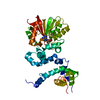
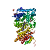
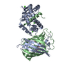
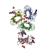
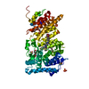
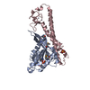
- PDB-4lil: Crystal structure of the catalytic subunit of human primase bound... -
+
Open data
ID or keywords:
Loading...
-
Basic information
Entry
Database: PDB / ID: 4lil
Title
Crystal structure of the catalytic subunit of human primase bound to UTP and Mn
Components
DNA primase small subunit
Keywords
TRANSFERASE / Prim fold
Function / homology
Function and homology information
ribonucleotide binding / DNA primase AEP / DNA replication initiation / Inhibition of replication initiation of damaged DNA by RB1/E2F1 / alpha DNA polymerase:primase complex / Telomere C-strand synthesis initiation / Polymerase switching / Processive synthesis on the lagging strand / Removal of the Flap Intermediate / DNA replication, synthesis of primer ...ribonucleotide binding / DNA primase AEP / DNA replication initiation / Inhibition of replication initiation of damaged DNA by RB1/E2F1 / alpha DNA polymerase:primase complex / Telomere C-strand synthesis initiation / Polymerase switching / Processive synthesis on the lagging strand / Removal of the Flap Intermediate / DNA replication, synthesis of primer / Polymerase switching on the C-strand of the telomere / Activation of the pre-replicative complex / DNA replication initiation / Defective pyroptosis / DNA-directed RNA polymerase activity / magnesium ion binding / zinc ion binding / nucleoplasm / membrane Similarity search - Function
DNA primase, PRIM domain / DNA primase, PRIM domain / DNA primase, small subunit, eukaryotic/archaeal / DNA primase, small subunit / DNA primase small subunit / Alpha-Beta Complex / Alpha Beta Similarity search - Domain/homology
In the structure databanks used in Yorodumi, some data are registered as the other names, "COVID-19 virus" and "2019-nCoV". Here are the details of the virus and the list of structure data.
Jan 31, 2019. EMDB accession codes are about to change! (news from PDBe EMDB page)
EMDB accession codes are about to change! (news from PDBe EMDB page)
The allocation of 4 digits for EMDB accession codes will soon come to an end. Whilst these codes will remain in use, new EMDB accession codes will include an additional digit and will expand incrementally as the available range of codes is exhausted. The current 4-digit format prefixed with “EMD-” (i.e. EMD-XXXX) will advance to a 5-digit format (i.e. EMD-XXXXX), and so on. It is currently estimated that the 4-digit codes will be depleted around Spring 2019, at which point the 5-digit format will come into force.
The EM Navigator/Yorodumi systems omit the EMD- prefix.
Related info.:Q: What is EMD? / ID/Accession-code notation in Yorodumi/EM Navigator
Yorodumi is a browser for structure data from EMDB, PDB, SASBDB, etc.
This page is also the successor to EM Navigator detail page, and also detail information page/front-end page for Omokage search.
The word "yorodu" (or yorozu) is an old Japanese word meaning "ten thousand". "mi" (miru) is to see.
Related info.:EMDB / PDB / SASBDB / Comparison of 3 databanks / Yorodumi Search / Aug 31, 2016. New EM Navigator & Yorodumi / Yorodumi Papers / Jmol/JSmol / Function and homology information / Changes in new EM Navigator and Yorodumi
 Movie
Movie Controller

 Yorodumi
 Open data
 Basic information
 Components
 Keywords
 Function and homology information
 Homo sapiens (human)
 X-RAY DIFFRACTION /
  Authors
 Citation
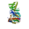
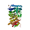
 Structure visualization
 Downloads & links
 Other downloads
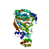


 PDBj
PDBj


 Assembly
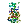


 Mass: 65.409 Da / Num. of mol.: 1 / Source method: obtained synthetically / Formula: Zn
 Mass: 54.938 Da / Num. of mol.: 2 / Source method: obtained synthetically / Formula: Mn
 Mass: 484.141 Da / Num. of mol.: 1 / Source method: obtained synthetically / Formula: C9H15N2O15P3 / Comment: UTP*YM
 Mass: 18.015 Da / Num. of mol.: 34 / Source method: isolated from a natural source / Formula: H2O
 Sample preparation
 / Beamline: 21-ID-F / Wavelength: 0.9787 Å
 Processing