 Movie
Movie Controller

[English] 日本語
 Yorodumi
- PDB-1r00: Crystal structure of aclacinomycin-10-hydroxylase (RdmB) in compl... -
+ Open data

- Basic information
| Entry | Database: PDB / ID: 1r00 | ||||||
|---|---|---|---|---|---|---|---|
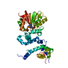
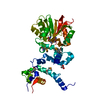
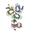
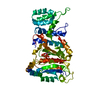
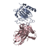
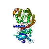
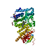
| Title | Crystal structure of aclacinomycin-10-hydroxylase (RdmB) in complex with S-adenosyl-L-homocysteine (SAH) | ||||||
 Components Components | aclacinomycin-10-hydroxylase | ||||||
 Keywords Keywords | OXIDOREDUCTASE / TRANSFERASE / Anthracycline / hydroxylase / methyltransferase / polyketide / Streptomyces / tailoring enzyme | ||||||
| Function / homology |  Function and homology information Function and homology informationLyases; Carbon-carbon lyases; Carboxy-lyases / carboxy-lyase activity / O-methyltransferase activity / antibiotic biosynthetic process / Transferases; Transferring one-carbon groups; Methyltransferases / methylation / protein dimerization activity Similarity search - Function | ||||||
| Biological species |  Streptomyces purpurascens (bacteria) Streptomyces purpurascens (bacteria) | ||||||
| Method |  X-RAY DIFFRACTION / SYNCHROTRON / fft / Resolution: 2.5 Å X-RAY DIFFRACTION / SYNCHROTRON / fft / Resolution: 2.5 Å | ||||||
 Authors Authors | Jansson, A. / Niemi, J. / Lindqvist, Y. / Mantsala, P. / Schneider, G. | ||||||
 Citation Citation | Journal: J.Mol.Biol. / Year: 2003 Title: Crystal Structure of Aclacinomycin-10-Hydroxylase, a S-Adenosyl-L-Methionine-dependent Methyltransferase Homolog Involved in Anthracycline Biosynthesis in Streptomyces purpurascens. Authors: Jansson, A. / Niemi, J. / Lindqvist, Y. / Mantsala, P. / Schneider, G. #1: Journal: Acta Crystallogr.,Sect.D / Year: 2003Title: Crystallization and preliminary X-ray diffraction studies of aclacinomycin-10-methylesterase and aclacinomycin-10-hydroxylase from Streptomyces purpurascens Authors: Jansson, A. / Niemi, J. / Mantsala, P. / Schneider, G. | ||||||
| History |
|
- Structure visualization
| Structure viewer | Molecule: MolmilJmol/JSmol |
|---|
- Downloads & links
-Download
| PDBx/mmCIF format | 1r00.cif.gz | 80.4 KB | Display | PDBx/mmCIF format |
|---|---|---|---|---|
| PDB format | pdb1r00.ent.gz | 59.3 KB | Display | PDB format |
| PDBx/mmJSON format | 1r00.json.gz | Tree view | PDBx/mmJSON format | |
| Others |  Other downloads Other downloads |
-Validation report
| Arichive directory | https://data.pdbj.org/pub/pdb/validation_reports/r0/1r00ftp://data.pdbj.org/pub/pdb/validation_reports/r0/1r00 | HTTPS FTP |
|---|
-Related structure data
| Related structure data |  1qzzSC S: Starting model for refinement C: citing same article ( |
|---|---|
| Similar structure data |

-Links
 PDBj
PDBj- Assembly
| Deposited unit | 
| ||||||||
|---|---|---|---|---|---|---|---|---|---|
| 1 |
| ||||||||
| 2 | 
| ||||||||
| Unit cell |
|
-Components
| #1: Protein | Mass: 39837.945 Da / Num. of mol.: 1 Source method: isolated from a genetically manipulated source Source: (gene. exp.) Streptomyces purpurascens (bacteria) / Gene: rdmb / Plasmid: pRDM16 / Production host: |
|---|---|
| #2: Chemical | ChemComp-ACT /   Mass: 59.044 Da / Num. of mol.: 1 / Source method: obtained synthetically / Formula: C2H3O2 Mass: 59.044 Da / Num. of mol.: 1 / Source method: obtained synthetically / Formula: C2H3O2 |
| #3: Chemical | ChemComp-SAH /   Type: L-peptide linking / Mass: 384.411 Da / Num. of mol.: 1 / Source method: obtained synthetically / Formula: C14H20N6O5S Type: L-peptide linking / Mass: 384.411 Da / Num. of mol.: 1 / Source method: obtained synthetically / Formula: C14H20N6O5S |
| #4: Water | ChemComp-HOH /  Mass: 18.015 Da / Num. of mol.: 91 / Source method: isolated from a natural source / Formula: H2O Mass: 18.015 Da / Num. of mol.: 91 / Source method: isolated from a natural source / Formula: H2O |
-Experimental details
-Experiment
| Experiment | Method: X-RAY DIFFRACTION / Number of used crystals: 1 |
|---|
- Sample preparation
| Crystal | Density Matthews: 2.1 Å3/Da / Density % sol: 41.52 % | ||||||||||||||||||||||||||||||
|---|---|---|---|---|---|---|---|---|---|---|---|---|---|---|---|---|---|---|---|---|---|---|---|---|---|---|---|---|---|---|---|
| Crystal grow | Temperature: 294 K / Method: vapor diffusion, hanging drop / pH: 5 Details: PEG4000, ammonium acetate, sodium acetate, pH 5.0, VAPOR DIFFUSION, HANGING DROP, temperature 294K | ||||||||||||||||||||||||||||||
| Crystal grow | *PLUS Method: vapor diffusion, hanging drop | ||||||||||||||||||||||||||||||
| Components of the solutions | *PLUS
|
-Data collection
| Diffraction | Mean temperature: 100 K |
|---|---|
| Diffraction source | Source: SYNCHROTRON / Site: MAX II  / Beamline: I711 / Wavelength: 1.076 Å / Beamline: I711 / Wavelength: 1.076 Å |
| Detector | Type: MARRESEARCH / Detector: CCD / Date: Feb 9, 2002 |
| Radiation | Protocol: SINGLE WAVELENGTH / Monochromatic (M) / Laue (L): M / Scattering type: x-ray |
| Radiation wavelength | Wavelength: 1.076 Å / Relative weight: 1 |
| Reflection | Resolution: 2.5→30 Å / Num. all: 12651 / Num. obs: 12651 / % possible obs: 99.8 % / Observed criterion σ(F): 0 / Observed criterion σ(I): 0 / Redundancy: 7.6 % / Biso Wilson estimate: 34 Å2 / Rsym value: 0.108 / Net I/σ(I): 10.4 |
| Reflection shell | Resolution: 2.5→2.59 Å / Mean I/σ(I) obs: 5.6 / Rsym value: 0.252 / % possible all: 99.8 |
| Reflection | *PLUS Num. measured all: 96318 / Rmerge(I) obs: 0.108 |
| Reflection shell | *PLUS Highest resolution: 2.5 Å / % possible obs: 99.8 % / Rmerge(I) obs: 0.252 |
- Processing
| Software |
| ||||||||||||||||||||
|---|---|---|---|---|---|---|---|---|---|---|---|---|---|---|---|---|---|---|---|---|---|
| Refinement | Method to determine structure: fft Starting model: PDB entry 1QZZ, SAM-complex Resolution: 2.5→30 Å Isotropic thermal model: Individual isotropic B-factors for each atom Cross valid method: THROUGHOUT / σ(F): 0 / Stereochemistry target values: maximum likelihood Details: riding hydrogens; the occupancy is put to zero on the sidechains of Gln15, Asp57, Lys84, Glu219, Arg298, Arg319
| ||||||||||||||||||||
| Displacement parameters | Biso mean: 32 Å2 | ||||||||||||||||||||
| Refinement step | Cycle: LAST / Resolution: 2.5→30 Å
| ||||||||||||||||||||
| Refine LS restraints |
| ||||||||||||||||||||
| LS refinement shell | Resolution: 2.5→2.59 Å /
| ||||||||||||||||||||
| Software | *PLUS Version: 5 / Classification: refinement | ||||||||||||||||||||
| Refinement | *PLUS % reflection Rfree: 9 % / Rfactor Rfree: 0.28 | ||||||||||||||||||||
| Solvent computation | *PLUS | ||||||||||||||||||||
| Displacement parameters | *PLUS | ||||||||||||||||||||
| Refine LS restraints | *PLUS
|
