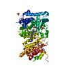
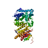
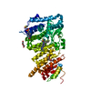
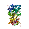
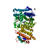
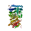
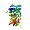
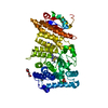
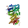
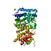
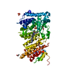
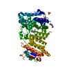
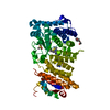
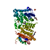
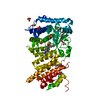
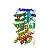
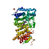
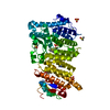
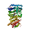
Entry Database : PDB / ID : 5db0Title Menin in complex with MI-352 Menin Keywords / / Function / homology Function Domain/homology Component
/ / / / / / / / / / / / / / / / / / / / / / / / / / / / / / / / / / / / / / / / / / / / / / / / / / / / / / / / / Biological species Homo sapiens (human)Method / / / Resolution : 1.5 Å Authors Pollock, J. / Dmitry, B. / Cierpicki, T. / Grembecka, J. Funding support Organization Grant number Country National Institutes of Health/National Cancer Institute (NIH/NCI) 1R)1CA160467 Leukemia & Lymphoma Society 6116-12 Leukemia & Lymphoma Society 1215-14 Leukemia & Lymphoma Society Therapy Accelerated Program American Chemical Society RSG-11-082-01-DMC American Chemical Society RSG-13-130-01-CDD APS LSCAT 085P1000817
Journal : J.Med.Chem. / Year : 2016Title : Property Focused Structure-Based Optimization of Small Molecule Inhibitors of the Protein-Protein Interaction between Menin and Mixed Lineage Leukemia (MLL).Authors : Borkin, D. / Pollock, J. / Kempinska, K. / Purohit, T. / Li, X. / Wen, B. / Zhao, T. / Miao, H. / Shukla, S. / He, M. / Sun, D. / Cierpicki, T. / Grembecka, J. History Deposition Aug 20, 2015 Deposition site / Processing site Revision 1.0 Mar 30, 2016 Provider / Type Revision 1.1 Sep 27, 2017 Group / Database references / Derived calculationsCategory / pdbx_audit_support / pdbx_struct_oper_listItem / _pdbx_audit_support.funding_organization / _pdbx_struct_oper_list.symmetry_operationRevision 1.2 Dec 4, 2019 Group / Category / Item Revision 1.3 Sep 27, 2023 Group Data collection / Database references ... Data collection / Database references / Derived calculations / Refinement description / Structure summary Category chem_comp / chem_comp_atom ... chem_comp / chem_comp_atom / chem_comp_bond / database_2 / entity / pdbx_entity_nonpoly / pdbx_initial_refinement_model Item _chem_comp.name / _database_2.pdbx_DOI ... _chem_comp.name / _database_2.pdbx_DOI / _database_2.pdbx_database_accession / _entity.pdbx_description / _pdbx_entity_nonpoly.name
Show all Show less
 Movie
Movie Controller


 Open data
 Basic information
 Components
 Keywords
 Function and homology information
 Homo sapiens (human)
 X-RAY DIFFRACTION /
  Authors
 United States, 7items
  Citation
 Structure visualization
 Downloads & links
 Other downloads
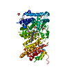


 PDBj
PDBj


 Assembly
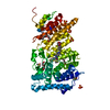


 Mass: 194.226 Da / Num. of mol.: 3 / Source method: obtained synthetically / Formula: C8H18O5 / Comment: precipitant*YM
 Mass: 96.063 Da / Num. of mol.: 4 / Source method: obtained synthetically / Formula: SO4
 Mass: 78.133 Da / Num. of mol.: 4 / Source method: obtained synthetically / Formula: C2H6OS / Comment: DMSO, precipitant*YM
 Mass: 544.592 Da / Num. of mol.: 1 / Source method: obtained synthetically / Formula: C26H27F3N6O2S
 Mass: 544.592 Da / Num. of mol.: 1 / Source method: obtained synthetically / Formula: C26H27F3N6O2S
 Sample preparation
 Processing