 Movie
Movie Controller


+ Open data

- Basic information
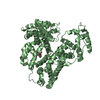
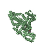
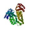
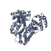
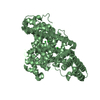
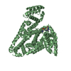
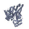
| Entry | Database: PDB / ID: 4lb2 | ||||||
|---|---|---|---|---|---|---|---|
| Title | X-ray study of human serum albumin complexed with idarubicin | ||||||
 Components Components | SERUM ALBUMIN | ||||||
 Keywords Keywords | TRANSPORT PROTEIN / PLASMA PROTEIN / CANCER / ONCOLOGY DRUG COMPLEX | ||||||
| Function / homology |  Function and homology information Function and homology informationexogenous protein binding / Ciprofloxacin ADME / enterobactin binding / cellular response to calcium ion starvation / Heme biosynthesis / HDL remodeling / negative regulation of mitochondrial depolarization / Prednisone ADME / Heme degradation / Aspirin ADME ...exogenous protein binding / Ciprofloxacin ADME / enterobactin binding / cellular response to calcium ion starvation / Heme biosynthesis / HDL remodeling / negative regulation of mitochondrial depolarization / Prednisone ADME / Heme degradation / Aspirin ADME / antioxidant activity / toxic substance binding / Scavenging of heme from plasma / Recycling of bile acids and salts / cellular response to starvation / platelet alpha granule lumen / fatty acid binding / Post-translational protein phosphorylation / Cytoprotection by HMOX1 / Regulation of Insulin-like Growth Factor (IGF) transport and uptake by Insulin-like Growth Factor Binding Proteins (IGFBPs) / Platelet degranulation / pyridoxal phosphate binding / protein-folding chaperone binding / blood microparticle / copper ion binding / endoplasmic reticulum lumen / Golgi apparatus / endoplasmic reticulum / protein-containing complex / DNA binding / extracellular space / extracellular exosome / extracellular region / identical protein binding / nucleus / cytoplasm Similarity search - Function | ||||||
| Biological species |  Homo sapiens (human) Homo sapiens (human) | ||||||
| Method |  X-RAY DIFFRACTION / MOLECULAR REPLACEMENT / Resolution: 2.8 Å X-RAY DIFFRACTION / MOLECULAR REPLACEMENT / Resolution: 2.8 Å | ||||||
 Authors Authors | Wang, Z. / Ho, J.X. / Ruble, J. / Rose, J.P. / Carter, D.C. | ||||||
 Citation Citation | Journal: Biochim.Biophys.Acta / Year: 2013 Title: Structural studies of several clinically important oncology drugs in complex with human serum albumin. Authors: Wang, Z.M. / Ho, J.X. / Ruble, J.R. / Rose, J. / Ruker, F. / Ellenburg, M. / Murphy, R. / Click, J. / Soistman, E. / Wilkerson, L. / Carter, D.C. #1: Journal: Burgers medicinal chemistry drug design and development, 7th editionYear: 2010 Title: Crystallographic survey of albumin drug interaction and preliminary applications in cancer chemotherapy Authors: Carter, D.C. #2: Journal: Adv.Protein Chem. / Year: 1994Title: Structure of Serum Albumin Authors: Carter, D.C. / Ho, J.X. #3: Journal: Eur.J.Biochem. / Year: 1994 Title: Preliminary crystallographic studies of four crystal forms of serum albumin. Authors: Carter, D.C. / Chang, B. / Ho, J.X. / Keeling, K. / Krishnasami, Z. #4: Journal: Nature / Year: 1993Title: Erratum. Atomic Structure and Chemistry of Human Serum Albumin Authors: Ho, X.M. / Carter, D.C. #5: Journal: Nature / Year: 1992Title: Atomic structure and chemistry of human serum albumin. Authors: He, X.M. / Carter, D.C. #6: Journal: Science / Year: 1990Title: Structure of Human Serum Albumin Authors: Carter, D.C. / Ho, X.M. #7: Journal: Science / Year: 1989 Title: Three-dimensional structure of human serum albumin. Authors: Carter, D.C. / He, X.M. / Munson, S.H. / Twigg, P.D. / Gernert, K.M. / Broom, M.B. / Miller, T.Y. | ||||||
| History |
|
- Structure visualization
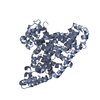
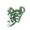
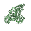
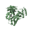
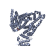
| Structure viewer | Molecule: MolmilJmol/JSmol |
|---|
- Downloads & links
-Download
| PDBx/mmCIF format | 4lb2.cif.gz | 234.3 KB | Display | PDBx/mmCIF format |
|---|---|---|---|---|
| PDB format | pdb4lb2.ent.gz | 196.8 KB | Display | PDB format |
| PDBx/mmJSON format | 4lb2.json.gz | Tree view | PDBx/mmJSON format | |
| Others |  Other downloads Other downloads |
-Validation report
| Summary document | 4lb2_validation.pdf.gz | 1.5 MB | Display | wwPDB validaton report |
|---|---|---|---|---|
| Full document | 4lb2_full_validation.pdf.gz | 1.5 MB | Display | |
| Data in XML | 4lb2_validation.xml.gz | 41.9 KB | Display | |
| Data in CIF | 4lb2_validation.cif.gz | 56.4 KB | Display | |
| Arichive directory | https://data.pdbj.org/pub/pdb/validation_reports/lb/4lb2ftp://data.pdbj.org/pub/pdb/validation_reports/lb/4lb2 | HTTPS FTP |
-Related structure data


-Links
 PDBj
PDBj


- Assembly
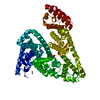
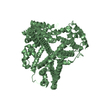
| Deposited unit | 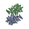
| ||||||||
|---|---|---|---|---|---|---|---|---|---|
| 1 | 
| ||||||||
| 2 | 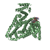
| ||||||||
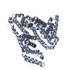
| Unit cell |
| ||||||||
| Noncrystallographic symmetry (NCS) | NCS domain: (Details: chain B and segid HSB) NCS domain segments: (Selection details: chain 'B' and segid 'HSB ') |
-Components
| #1: Protein | Mass: 66571.219 Da / Num. of mol.: 2 / Fragment: UNP residues 25-609 / Source method: isolated from a natural source / Source: (natural) Homo sapiens (human) / References: UniProt: P02768#2: Chemical | ChemComp-DM5 /   Mass: 497.494 Da / Num. of mol.: 4 / Source method: obtained synthetically / Formula: C26H27NO9 Mass: 497.494 Da / Num. of mol.: 4 / Source method: obtained synthetically / Formula: C26H27NO9#3: Water | ChemComp-HOH / |  Mass: 18.015 Da / Num. of mol.: 75 / Source method: isolated from a natural source / Formula: H2O Mass: 18.015 Da / Num. of mol.: 75 / Source method: isolated from a natural source / Formula: H2O |
|---|
-Experimental details
-Experiment
| Experiment | Method: X-RAY DIFFRACTION / Number of used crystals: 1 |
|---|
- Sample preparation
| Crystal | Density Matthews: 2.28 Å3/Da / Density % sol: 46.11 % |
|---|---|
| Crystal grow | Temperature: 293 K / Method: vapor diffusion, sitting drop / pH: 7.5 Details: PEG 3350, POTASSIUM PHOSPHATE, Crystals of the complexes were obtained by standard vapor equilibration methods with conditions optimized by screens varying protein concentration, pH, drug ...Details: PEG 3350, POTASSIUM PHOSPHATE, Crystals of the complexes were obtained by standard vapor equilibration methods with conditions optimized by screens varying protein concentration, pH, drug molar ratios, centered on the original crystallization hit using protocols described previously for the monoclinic [Carter, et al., Eur. J. Biochemistry (1994) 226: 1049-1052] and triclinic [Sugo, et al., Protein Eng (1999) 12: 439-446] crystal forms., pH 7.5, vapor diffusion, SITTING DROP, temperature 293K |
-Data collection
| Diffraction | Mean temperature: 100 K | ||||||||||||||||||||||||||||||||||||||||||||||||||||||||||||||||||
|---|---|---|---|---|---|---|---|---|---|---|---|---|---|---|---|---|---|---|---|---|---|---|---|---|---|---|---|---|---|---|---|---|---|---|---|---|---|---|---|---|---|---|---|---|---|---|---|---|---|---|---|---|---|---|---|---|---|---|---|---|---|---|---|---|---|---|---|
| Diffraction source | Source: ROTATING ANODE / Type: RIGAKU RU300 / Wavelength: 1.5418 Å | ||||||||||||||||||||||||||||||||||||||||||||||||||||||||||||||||||
| Detector | Type: RIGAKU RAXIS IV / Detector: IMAGE PLATE / Date: Jan 1, 2003 / Details: CONFOCAL OPTICS | ||||||||||||||||||||||||||||||||||||||||||||||||||||||||||||||||||
| Radiation | Monochromator: CONFOCAL OPTICS / Protocol: SINGLE WAVELENGTH / Monochromatic (M) / Laue (L): M / Scattering type: x-ray | ||||||||||||||||||||||||||||||||||||||||||||||||||||||||||||||||||
| Radiation wavelength | Wavelength: 1.5418 Å / Relative weight: 1 | ||||||||||||||||||||||||||||||||||||||||||||||||||||||||||||||||||
| Reflection | Resolution: 2.7→30 Å / Num. obs: 31105 / % possible obs: 96.2 % / Rmerge(I) obs: 0.075 / Χ2: 4.351 / Net I/σ(I): 13.8 | ||||||||||||||||||||||||||||||||||||||||||||||||||||||||||||||||||
| Reflection shell |
|
- Processing
| Software |
| |||||||||||||||||||||||||||||||||||||||||||||||||||||||||||||||||||||||||||||
|---|---|---|---|---|---|---|---|---|---|---|---|---|---|---|---|---|---|---|---|---|---|---|---|---|---|---|---|---|---|---|---|---|---|---|---|---|---|---|---|---|---|---|---|---|---|---|---|---|---|---|---|---|---|---|---|---|---|---|---|---|---|---|---|---|---|---|---|---|---|---|---|---|---|---|---|---|---|---|
| Refinement | Method to determine structure: MOLECULAR REPLACEMENT / Resolution: 2.8→28.163 Å / Occupancy max: 1 / Occupancy min: 1 / SU ML: 0.3 / σ(F): 1.97 / Phase error: 26.37 / Stereochemistry target values: ML
| |||||||||||||||||||||||||||||||||||||||||||||||||||||||||||||||||||||||||||||
| Solvent computation | Shrinkage radii: 0.9 Å / VDW probe radii: 1.11 Å / Solvent model: FLAT BULK SOLVENT MODEL | |||||||||||||||||||||||||||||||||||||||||||||||||||||||||||||||||||||||||||||
| Displacement parameters | Biso max: 103.23 Å2 / Biso mean: 43.1098 Å2 / Biso min: 12.8 Å2 | |||||||||||||||||||||||||||||||||||||||||||||||||||||||||||||||||||||||||||||
| Refinement step | Cycle: LAST / Resolution: 2.8→28.163 Å
| |||||||||||||||||||||||||||||||||||||||||||||||||||||||||||||||||||||||||||||
| Refine LS restraints |
| |||||||||||||||||||||||||||||||||||||||||||||||||||||||||||||||||||||||||||||
| Refine LS restraints NCS | Number: 5405 / Type: POSITIONAL / Rms dev position: 12.635 Å | |||||||||||||||||||||||||||||||||||||||||||||||||||||||||||||||||||||||||||||
| LS refinement shell | Refine-ID: X-RAY DIFFRACTION / Total num. of bins used: 10
|
