 Movie
Movie Controller


+ Open data

- Basic information
| Entry | Database: PDB / ID: 4jvw | ||||||
|---|---|---|---|---|---|---|---|
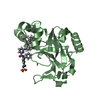
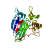
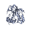
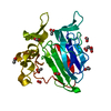
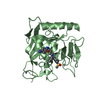
| Title | IgM C4-domain from mouse | ||||||
 Components Components | Ig mu chain C region secreted form | ||||||
 Keywords Keywords | PROTEIN BINDING / Immunoglobulin M / antibody / oligomerization / Immunoglobulin fold / Receptor | ||||||
| Function / homology |  Function and homology information Function and homology informationCD22 mediated BCR regulation / hexameric IgM immunoglobulin complex / IgM B cell receptor complex / pentameric IgM immunoglobulin complex / B cell receptor complex / Cell surface interactions at the vascular wall / regulation of immunoglobulin production / positive regulation of B cell activation / Antigen activates B Cell Receptor (BCR) leading to generation of second messengers / humoral immune response mediated by circulating immunoglobulin ...CD22 mediated BCR regulation / hexameric IgM immunoglobulin complex / IgM B cell receptor complex / pentameric IgM immunoglobulin complex / B cell receptor complex / Cell surface interactions at the vascular wall / regulation of immunoglobulin production / positive regulation of B cell activation / Antigen activates B Cell Receptor (BCR) leading to generation of second messengers / humoral immune response mediated by circulating immunoglobulin / pre-B cell allelic exclusion / early endosome to late endosome transport / B cell affinity maturation / regulation of cell morphogenesis / antigen processing and presentation / immunoglobulin complex, circulating / immunoglobulin receptor binding / immunoglobulin mediated immune response / complement activation, classical pathway / positive regulation of endocytosis / antigen binding / positive regulation of B cell proliferation / B cell receptor signaling pathway / positive regulation of immune response / transmembrane signaling receptor activity / antibacterial humoral response / defense response to Gram-negative bacterium / adaptive immune response / positive regulation of MAPK cascade / external side of plasma membrane / innate immune response / perinuclear region of cytoplasm / cell surface / : / membrane / identical protein binding / plasma membrane / cytoplasm Similarity search - Function | ||||||
| Biological species |  | ||||||
| Method |  X-RAY DIFFRACTION / MOLECULAR REPLACEMENT / Resolution: 2 Å X-RAY DIFFRACTION / MOLECULAR REPLACEMENT / Resolution: 2 Å | ||||||
 Authors Authors | Mueller, R. / Graewert, A.M. / Kern, T. / Madl, T. / Peschek, J. / Sattler, M. / Groll, M. / Buchner, J. | ||||||
 Citation Citation | Journal: Proc.Natl.Acad.Sci.USA / Year: 2013 Title: High-resolution structures of the IgM Fc domains reveal principles of its hexamer formation. Authors: Muller, R. / Grawert, M.A. / Kern, T. / Madl, T. / Peschek, J. / Sattler, M. / Groll, M. / Buchner, J. | ||||||
| History |
|
- Structure visualization
| Structure viewer | Molecule: MolmilJmol/JSmol |
|---|
- Downloads & links
-Download
| PDBx/mmCIF format | 4jvw.cif.gz | 183.7 KB | Display | PDBx/mmCIF format |
|---|---|---|---|---|
| PDB format | pdb4jvw.ent.gz | 147.9 KB | Display | PDB format |
| PDBx/mmJSON format | 4jvw.json.gz | Tree view | PDBx/mmJSON format | |
| Others |  Other downloads Other downloads |
-Validation report
| Arichive directory | https://data.pdbj.org/pub/pdb/validation_reports/jv/4jvwftp://data.pdbj.org/pub/pdb/validation_reports/jv/4jvw | HTTPS FTP |
|---|
-Related structure data
| Related structure data |  4ba8C  4jvuC 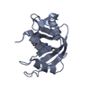 1jvuS C: citing same article ( S: Starting model for refinement |
|---|---|
| Similar structure data |


-Links
 PDBj
PDBj


- Assembly
| Deposited unit | 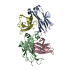
| |||||||||||||||||||||||||||||||||||||||||||||||||||||||||||||||||
|---|---|---|---|---|---|---|---|---|---|---|---|---|---|---|---|---|---|---|---|---|---|---|---|---|---|---|---|---|---|---|---|---|---|---|---|---|---|---|---|---|---|---|---|---|---|---|---|---|---|---|---|---|---|---|---|---|---|---|---|---|---|---|---|---|---|---|
| 1 | 
| |||||||||||||||||||||||||||||||||||||||||||||||||||||||||||||||||
| 2 | 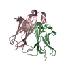
| |||||||||||||||||||||||||||||||||||||||||||||||||||||||||||||||||
| Unit cell |
| |||||||||||||||||||||||||||||||||||||||||||||||||||||||||||||||||
| Components on special symmetry positions |
| |||||||||||||||||||||||||||||||||||||||||||||||||||||||||||||||||
| Noncrystallographic symmetry (NCS) | NCS domain:
NCS domain segments:
NCS oper:
|
-Components
| #1: Protein | Mass: 12743.415 Da / Num. of mol.: 4 / Fragment: uno residues 324-436 Source method: isolated from a genetically manipulated source Source: (gene. exp.)  #2: Water | ChemComp-HOH / |  Mass: 18.015 Da / Num. of mol.: 428 / Source method: isolated from a natural source / Formula: H2O Mass: 18.015 Da / Num. of mol.: 428 / Source method: isolated from a natural source / Formula: H2OHas protein modification | Y | |
|---|
-Experimental details
-Experiment
| Experiment | Method: X-RAY DIFFRACTION / Number of used crystals: 1 |
|---|
- Sample preparation
| Crystal | Density Matthews: 2.29 Å3/Da / Density % sol: 46.34 % |
|---|---|
| Crystal grow | Temperature: 293 K / Method: vapor diffusion, hanging drop / pH: 7.2 Details: 0.1 M HEPES, 15% PEG 8000, pH 7.2, VAPOR DIFFUSION, HANGING DROP, temperature 293K |
-Data collection
| Diffraction | Mean temperature: 100 K |
|---|---|
| Diffraction source | Source: ROTATING ANODE / Type: BRUKER AXS MICROSTAR / Wavelength: 1.5418 Å |
| Detector | Type: Bruker Platinum 135 / Detector: CCD / Date: Feb 15, 2009 |
| Radiation | Monochromator: Scanning emission monochromator / Protocol: SINGLE WAVELENGTH / Monochromatic (M) / Laue (L): M / Scattering type: x-ray |
| Radiation wavelength | Wavelength: 1.5418 Å / Relative weight: 1 |
| Reflection | Resolution: 2→40 Å / Num. all: 31721 / Num. obs: 31721 / % possible obs: 100 % / Observed criterion σ(F): 2 / Observed criterion σ(I): 2 / Redundancy: 4.9 % / Rmerge(I) obs: 0.046 / Net I/σ(I): 19 |
| Reflection shell | Resolution: 2→2.1 Å / Rmerge(I) obs: 0.354 / Mean I/σ(I) obs: 3.1 / % possible all: 100 |
- Processing
| Software |
| |||||||||||||||||||||||||||||||||||||||||||||||||||||||||||||||||||||||||||||||||||||||||||||||||||||||||||||||||||||||||||||
|---|---|---|---|---|---|---|---|---|---|---|---|---|---|---|---|---|---|---|---|---|---|---|---|---|---|---|---|---|---|---|---|---|---|---|---|---|---|---|---|---|---|---|---|---|---|---|---|---|---|---|---|---|---|---|---|---|---|---|---|---|---|---|---|---|---|---|---|---|---|---|---|---|---|---|---|---|---|---|---|---|---|---|---|---|---|---|---|---|---|---|---|---|---|---|---|---|---|---|---|---|---|---|---|---|---|---|---|---|---|---|---|---|---|---|---|---|---|---|---|---|---|---|---|---|---|---|
| Refinement | Method to determine structure: MOLECULAR REPLACEMENT Starting model: 1JVU Resolution: 2→15 Å / Cor.coef. Fo:Fc: 0.948 / Cor.coef. Fo:Fc free: 0.926 / SU B: 8.441 / SU ML: 0.123 / Cross valid method: THROUGHOUT / ESU R: 0.217 / ESU R Free: 0.176 / Stereochemistry target values: MAXIMUM LIKELIHOOD / Details: HYDROGENS HAVE BEEN ADDED IN THE RIDING POSITIONS
| |||||||||||||||||||||||||||||||||||||||||||||||||||||||||||||||||||||||||||||||||||||||||||||||||||||||||||||||||||||||||||||
| Solvent computation | Ion probe radii: 0.8 Å / Shrinkage radii: 0.8 Å / VDW probe radii: 1.2 Å / Solvent model: MASK | |||||||||||||||||||||||||||||||||||||||||||||||||||||||||||||||||||||||||||||||||||||||||||||||||||||||||||||||||||||||||||||
| Displacement parameters | Biso mean: 26.083 Å2
| |||||||||||||||||||||||||||||||||||||||||||||||||||||||||||||||||||||||||||||||||||||||||||||||||||||||||||||||||||||||||||||
| Refinement step | Cycle: LAST / Resolution: 2→15 Å
| |||||||||||||||||||||||||||||||||||||||||||||||||||||||||||||||||||||||||||||||||||||||||||||||||||||||||||||||||||||||||||||
| Refine LS restraints |
| |||||||||||||||||||||||||||||||||||||||||||||||||||||||||||||||||||||||||||||||||||||||||||||||||||||||||||||||||||||||||||||
| Refine LS restraints NCS | Ens-ID: 1 / Number: 1577 / Refine-ID: X-RAY DIFFRACTION
| |||||||||||||||||||||||||||||||||||||||||||||||||||||||||||||||||||||||||||||||||||||||||||||||||||||||||||||||||||||||||||||
| LS refinement shell | Resolution: 2→2.051 Å / Total num. of bins used: 20
| |||||||||||||||||||||||||||||||||||||||||||||||||||||||||||||||||||||||||||||||||||||||||||||||||||||||||||||||||||||||||||||
| Refinement TLS params. | Method: refined / Refine-ID: X-RAY DIFFRACTION
| |||||||||||||||||||||||||||||||||||||||||||||||||||||||||||||||||||||||||||||||||||||||||||||||||||||||||||||||||||||||||||||
| Refinement TLS group |
|
