 Movie
Movie Controller


+ Open data

- Basic information
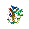
| Entry | Database: PDB / ID: 4h0g | ||||||
|---|---|---|---|---|---|---|---|
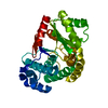
| Title | Crystal structure of mimicry-recognizing native 2D10 scFv | ||||||
 Components Components | 2D10 scFv | ||||||
 Keywords Keywords | IMMUNE SYSTEM / molecular mimicry / antigen binding / sugar | ||||||
| Function / homology | Immunoglobulins / Immunoglobulin-like / Sandwich / Mainly Beta Function and homology information Function and homology information | ||||||
| Biological species |  | ||||||
| Method |  X-RAY DIFFRACTION / MOLECULAR REPLACEMENT / Resolution: 2.3 Å X-RAY DIFFRACTION / MOLECULAR REPLACEMENT / Resolution: 2.3 Å | ||||||
 Authors Authors | Tapryal, S. / Gaur, V. / Kaur, K.J. / Salunke, D.M. | ||||||
 Citation Citation | Journal: J.Immunol. / Year: 2013 Title: Structural evaluation of a mimicry-recognizing paratope: plasticity in antigen-antibody interactions manifests in molecular mimicry. Authors: Tapryal, S. / Gaur, V. / Kaur, K.J. / Salunke, D.M. | ||||||
| History |
|
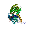
- Structure visualization
| Structure viewer | Molecule: MolmilJmol/JSmol |
|---|
- Downloads & links
-Download
| PDBx/mmCIF format | 4h0g.cif.gz | 63.2 KB | Display | PDBx/mmCIF format |
|---|---|---|---|---|
| PDB format | pdb4h0g.ent.gz | 46.1 KB | Display | PDB format |
| PDBx/mmJSON format | 4h0g.json.gz | Tree view | PDBx/mmJSON format | |
| Others |  Other downloads Other downloads |
-Validation report
| Arichive directory | https://data.pdbj.org/pub/pdb/validation_reports/h0/4h0gftp://data.pdbj.org/pub/pdb/validation_reports/h0/4h0g | HTTPS FTP |
|---|
-Related structure data
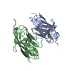
| Related structure data |  4h0hSC 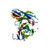 4h0iC S: Starting model for refinement C: citing same article ( |
|---|---|
| Similar structure data |


-Links
 PDBj
PDBj

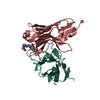
- Assembly
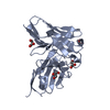
| Deposited unit | 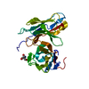
| ||||||||
|---|---|---|---|---|---|---|---|---|---|
| 1 |
| ||||||||
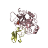
| Unit cell |
|
-Components
| #1: Antibody | Mass: 26894.973 Da / Num. of mol.: 1 Source method: isolated from a genetically manipulated source Source: (gene. exp.)  |
|---|---|
| #2: Chemical | ChemComp-TRS /   Mass: 122.143 Da / Num. of mol.: 1 / Source method: obtained synthetically / Formula: C4H12NO3 / Comment: pH buffer*YM Mass: 122.143 Da / Num. of mol.: 1 / Source method: obtained synthetically / Formula: C4H12NO3 / Comment: pH buffer*YM |
| #3: Water | ChemComp-HOH /  Mass: 18.015 Da / Num. of mol.: 176 / Source method: isolated from a natural source / Formula: H2O Mass: 18.015 Da / Num. of mol.: 176 / Source method: isolated from a natural source / Formula: H2O |
| Has protein modification | Y |
-Experimental details
-Experiment
| Experiment | Method: X-RAY DIFFRACTION / Number of used crystals: 1 |
|---|
- Sample preparation
| Crystal | Density Matthews: 2.48 Å3/Da / Density % sol: 50.44 % |
|---|---|
| Crystal grow | Temperature: 277 K / Method: vapor diffusion, hanging drop / pH: 6.5 Details: 50 mM MES, pH 6.5, 1.6 M magnesium sulfate, VAPOR DIFFUSION, HANGING DROP, temperature 277K |
-Data collection
| Diffraction | Mean temperature: 120 K |
|---|---|
| Diffraction source | Source: ROTATING ANODE / Type: RIGAKU RU300 / Wavelength: 1.5418 Å |
| Detector | Type: MAR scanner 345 mm plate / Detector: IMAGE PLATE / Date: Apr 9, 2009 / Details: mirrors |
| Radiation | Monochromator: Ni filter CMF 12 38CU-6 (Osmic Inc.) / Protocol: SINGLE WAVELENGTH / Monochromatic (M) / Laue (L): M / Scattering type: x-ray |
| Radiation wavelength | Wavelength: 1.5418 Å / Relative weight: 1 |
| Reflection | Resolution: 2.2→35 Å / Num. all: 12529 / Num. obs: 12216 / % possible obs: 100 % / Redundancy: 14.1 % / Rmerge(I) obs: 0.077 / Net I/σ(I): 26.3 |
| Reflection shell | Resolution: 2.2→2.32 Å / Redundancy: 14 % / Rmerge(I) obs: 0.597 / Mean I/σ(I) obs: 4.6 / % possible all: 100 |
- Processing
| Software |
| ||||||||||||||||||||||||
|---|---|---|---|---|---|---|---|---|---|---|---|---|---|---|---|---|---|---|---|---|---|---|---|---|---|
| Refinement | Method to determine structure: MOLECULAR REPLACEMENT Starting model: PDB ENTRY 4H0H Resolution: 2.3→35 Å / Occupancy max: 1 / Occupancy min: 1 / σ(F): 0
| ||||||||||||||||||||||||
| Solvent computation | Bsol: 71.3332 Å2 | ||||||||||||||||||||||||
| Displacement parameters | Biso max: 136.55 Å2 / Biso mean: 44.5573 Å2 / Biso min: 14.09 Å2
| ||||||||||||||||||||||||
| Refinement step | Cycle: LAST / Resolution: 2.3→35 Å
| ||||||||||||||||||||||||
| Refine LS restraints |
| ||||||||||||||||||||||||
| LS refinement shell | Resolution: 2.2→2.25 Å
| ||||||||||||||||||||||||
| Xplor file |
|
