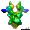
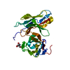
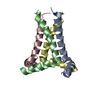
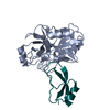
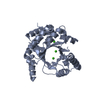
Journal: Proc Natl Acad Sci U S A / Year: 2013 Title: Structure and assembly of an inner membrane platform for initiation of type IV pilus biogenesis. Authors: Vijaykumar Karuppiah / Richard F Collins / Angela Thistlethwaite / Ya Gao / Jeremy P Derrick / Abstract: Type IV pili are long fibers that are assembled by polymerization of a major pilin protein in the periplasm of a wide range of bacteria and archaea. They play crucial roles in pathogenesis, DNA ...Type IV pili are long fibers that are assembled by polymerization of a major pilin protein in the periplasm of a wide range of bacteria and archaea. They play crucial roles in pathogenesis, DNA transformation, and motility, and are capable of rapid retraction, generating powerful motor forces. PilN and PilO are integral inner membrane proteins that are essential for type IV pilus formation. Here, we show that PilN and PilO from Thermus thermophilus can be isolated as a complex with PilM, a cytoplasmic protein with structural similarities to the cytoskeletal protein MreB. The crystal structure of the periplasmic portion of PilN forms a homodimer with an extensive, conserved interaction interface. We conducted serial 3D reconstructions by electron microscopy of PilMN, PilMNO, and PilMNO bound to the major pilin protein PilA4, to chart the assembly of the inner membrane pilus biogenesis platform. PilN drives the dimerization of the PilMN complex with a stoichiometry of 2:2; binding of two PilO monomers then causes the PilN periplasmic domains to dissociate. Finally, two PilA4 monomers bind to the periplasmic domains of PilN and PilO, to generate a T-shaped complex that is primed for addition of the pilin to the nascent pilus fiber. Docking of structures for PilM, PilN, PilO, and PilA4 into the electron density maps of the transmembrane complexes was used to generate a sequence of molecular structures that chart the initial events in type IV pilus formation, and provide structural information on the early events in this important secretion process.
Mass: 18.015 Da / Num. of mol.: 142 / Source method: isolated from a natural source / Formula: H2O
Sequence details
CYTOPLASMIC AND TRANSMEMBRANE REGIONS REMOVED. THE FIRST FOUR RESIDUES (GSHM) AT THE N-TERMINUS AND ...CYTOPLASMIC AND TRANSMEMBRANE REGIONS REMOVED. THE FIRST FOUR RESIDUES (GSHM) AT THE N-TERMINUS AND THE LAST EIGHT RESIDUES (LEHHHHHH) ARE FROM THE EXPRESSION VECTOR
-
Experimental details
-
Experiment
Experiment
Method: X-RAY DIFFRACTION / Number of used crystals: 1
-
Sample preparation
Crystal
Density Matthews: 1.8 Å3/Da / Density % sol: 32 % / Description: NONE
Crystal grow
pH: 7 Details: 0.1 M HEPES PH 7.0 0.2 M MAGNESIUM CHLORIDE 20% PEG 6000 10 MM ZINC CHLORIDE
Protocol: SINGLE WAVELENGTH / Monochromatic (M) / Laue (L): M / Scattering type: x-ray
Radiation wavelength
Wavelength: 0.969 Å / Relative weight: 1
Reflection
Resolution: 2.05→40 Å / Num. obs: 20669 / % possible obs: 99.4 % / Observed criterion σ(I): 2 / Redundancy: 3.4 % / Rmerge(I) obs: 0.07 / Net I/σ(I): 12.9
Reflection shell
Resolution: 2.05→2.1 Å / Redundancy: 3.4 % / Rmerge(I) obs: 0.57 / Mean I/σ(I) obs: 2.4 / % possible all: 98.8
-
Processing
Software
Name
Version
Classification
REFMAC
5.6.0117
refinement
XDS
datareduction
PHENIX
phasing
Refinement
Method to determine structure: SAD Starting model: NONE Resolution: 2.05→47.96 Å / Cor.coef. Fo:Fc: 0.945 / Cor.coef. Fo:Fc free: 0.928 / SU B: 3.6 / SU ML: 0.099 / Cross valid method: THROUGHOUT / ESU R: 0.158 / ESU R Free: 0.145 / Stereochemistry target values: MAXIMUM LIKELIHOOD / Details: HYDROGENS HAVE BEEN ADDED IN THE RIDING POSITIONS.
Rfactor
Num. reflection
% reflection
Selection details
Rfree
0.22623
1058
5.1 %
RANDOM
Rwork
0.19584
-
-
-
obs
0.19733
19607
99.38 %
-
Solvent computation
Ion probe radii: 0.8 Å / Shrinkage radii: 0.8 Å / VDW probe radii: 1.2 Å / Solvent model: MASK
 Movie
Movie Controller

 Yorodumi
 Open data
 Basic information
 Components
 Keywords
 Function and homology information
 THERMUS THERMOPHILUS (bacteria)
 X-RAY DIFFRACTION /
  Authors
 Citation
 Structure visualization
 Downloads & links
 Other downloads


 PDBj
PDBj Assembly

 Mass: 24.305 Da / Num. of mol.: 1 / Source method: obtained synthetically / Formula: Mg
 Mass: 18.015 Da / Num. of mol.: 142 / Source method: isolated from a natural source / Formula: H2O
 Sample preparation
 Processing