 Movie
Movie Controller

[English] 日本語
 Yorodumi
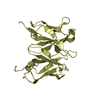
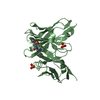
- PDB-4h0i: Crystal Structure of Scfv-2D10 in Complex with Methyl Alpha-D-Man... -
+ Open data

- Basic information
| Entry | Database: PDB / ID: 4h0i | ||||||
|---|---|---|---|---|---|---|---|
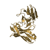
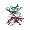
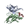
| Title | Crystal Structure of Scfv-2D10 in Complex with Methyl Alpha-D-Mannopyranoside | ||||||
 Components Components | 2D10 scFv | ||||||
 Keywords Keywords | IMMUNE SYSTEM / single chain antibody / molecular mimicry / antigen recognition | ||||||
| Function / homology | Immunoglobulins / Immunoglobulin-like / Sandwich / Mainly Beta / methyl alpha-D-mannopyranoside Function and homology information Function and homology information | ||||||
| Biological species |  | ||||||
| Method |  X-RAY DIFFRACTION / MOLECULAR REPLACEMENT / Resolution: 2.4 Å X-RAY DIFFRACTION / MOLECULAR REPLACEMENT / Resolution: 2.4 Å | ||||||
 Authors Authors | Tapryal, S. / Gaur, V. / Kaur, K.J. / Salunke, D.M. | ||||||
 Citation Citation | Journal: J.Immunol. / Year: 2013 Title: Structural evaluation of a mimicry-recognizing paratope: plasticity in antigen-antibody interactions manifests in molecular mimicry. Authors: Tapryal, S. / Gaur, V. / Kaur, K.J. / Salunke, D.M. | ||||||
| History |
|
- Structure visualization
| Structure viewer | Molecule: MolmilJmol/JSmol |
|---|
- Downloads & links
-Download
| PDBx/mmCIF format | 4h0i.cif.gz | 58.7 KB | Display | PDBx/mmCIF format |
|---|---|---|---|---|
| PDB format | pdb4h0i.ent.gz | 42.6 KB | Display | PDB format |
| PDBx/mmJSON format | 4h0i.json.gz | Tree view | PDBx/mmJSON format | |
| Others |  Other downloads Other downloads |
-Validation report
| Arichive directory | https://data.pdbj.org/pub/pdb/validation_reports/h0/4h0iftp://data.pdbj.org/pub/pdb/validation_reports/h0/4h0i | HTTPS FTP |
|---|
-Related structure data
| Related structure data |  4h0gC  4h0hC 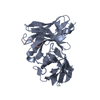 1jp5S C: citing same article ( S: Starting model for refinement |
|---|---|
| Similar structure data |


-Links
 PDBj
PDBj

- Assembly
| Deposited unit | 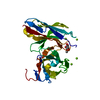
| ||||||||
|---|---|---|---|---|---|---|---|---|---|
| 1 |
| ||||||||
| Unit cell |
| ||||||||
| Components on special symmetry positions |
|
-Components
| #1: Antibody | Mass: 26894.973 Da / Num. of mol.: 1 Source method: isolated from a genetically manipulated source Source: (gene. exp.)  | ||||
|---|---|---|---|---|---|
| #2: Sugar | ChemComp-MMA / 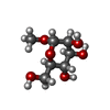  Type: D-saccharide / Mass: 194.182 Da / Num. of mol.: 1 Type: D-saccharide / Mass: 194.182 Da / Num. of mol.: 1Source method: isolated from a genetically manipulated source Formula: C7H14O6 | ||||
| #3: Chemical | ChemComp-MG /   Mass: 24.305 Da / Num. of mol.: 4 / Source method: obtained synthetically / Formula: Mg Mass: 24.305 Da / Num. of mol.: 4 / Source method: obtained synthetically / Formula: Mg#4: Water | ChemComp-HOH / |  Mass: 18.015 Da / Num. of mol.: 133 / Source method: isolated from a natural source / Formula: H2O Mass: 18.015 Da / Num. of mol.: 133 / Source method: isolated from a natural source / Formula: H2OHas protein modification | Y | |
-Experimental details
-Experiment
| Experiment | Method: X-RAY DIFFRACTION / Number of used crystals: 1 |
|---|
- Sample preparation
| Crystal | Density Matthews: 2.6 Å3/Da / Density % sol: 52.64 % |
|---|---|
| Crystal grow | Temperature: 277 K / Method: vapor diffusion, hanging drop / pH: 6.5 Details: 100 mM MES, pH 6.5, 1.6 M magnesium sulfate, VAPOR DIFFUSION, HANGING DROP, temperature 277K |
-Data collection
| Diffraction | Mean temperature: 120 K |
|---|---|
| Diffraction source | Source: ROTATING ANODE / Type: RIGAKU RU300 / Wavelength: 1.5418 |
| Detector | Type: MAR scanner 345 mm plate / Detector: IMAGE PLATE / Date: Apr 29, 2008 / Details: mirrors |
| Radiation | Monochromator: Ni filter CMF 12 38CU-6 (Osmic Inc.) / Protocol: SINGLE WAVELENGTH / Monochromatic (M) / Laue (L): M / Scattering type: x-ray |
| Radiation wavelength | Wavelength: 1.5418 Å / Relative weight: 1 |
| Reflection | Resolution: 2.4→69.516 Å / Num. obs: 11290 / % possible obs: 100 % / Observed criterion σ(I): 0 / Redundancy: 9.2 % / Biso Wilson estimate: 30.4 Å2 / Rmerge(I) obs: 0.13 / Net I/σ(I): 15.9 |
| Reflection shell | Resolution: 2.4→2.53 Å / Redundancy: 9.2 % / Rmerge(I) obs: 0.67 / Mean I/σ(I) obs: 3.2 / % possible all: 100 |
- Processing
| Software |
| ||||||||||||||||||||||||||||||||||||||||||||||||||||||||||||
|---|---|---|---|---|---|---|---|---|---|---|---|---|---|---|---|---|---|---|---|---|---|---|---|---|---|---|---|---|---|---|---|---|---|---|---|---|---|---|---|---|---|---|---|---|---|---|---|---|---|---|---|---|---|---|---|---|---|---|---|---|---|
| Refinement | Method to determine structure: MOLECULAR REPLACEMENT Starting model: PDB ENTRY 1JP5 Resolution: 2.4→34 Å / Rfactor Rfree error: 0.007 / Data cutoff high absF: 10000 / Data cutoff low absF: 0 / Isotropic thermal model: RESTRAINED / Cross valid method: THROUGHOUT / σ(F): 0
| ||||||||||||||||||||||||||||||||||||||||||||||||||||||||||||
| Solvent computation | Solvent model: FLAT MODEL / Bsol: 35.1527 Å2 / ksol: 0.38 e/Å3 | ||||||||||||||||||||||||||||||||||||||||||||||||||||||||||||
| Displacement parameters | Biso mean: 33.8 Å2
| ||||||||||||||||||||||||||||||||||||||||||||||||||||||||||||
| Refine analyze |
| ||||||||||||||||||||||||||||||||||||||||||||||||||||||||||||
| Refinement step | Cycle: LAST / Resolution: 2.4→34 Å
| ||||||||||||||||||||||||||||||||||||||||||||||||||||||||||||
| Refine LS restraints |
| ||||||||||||||||||||||||||||||||||||||||||||||||||||||||||||
| LS refinement shell | Resolution: 2.4→2.55 Å / Rfactor Rfree error: 0.022 / Total num. of bins used: 6
| ||||||||||||||||||||||||||||||||||||||||||||||||||||||||||||
| Xplor file |
|
