 Movie
Movie Controller

[English] 日本語
 Yorodumi
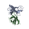
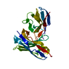
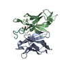
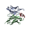
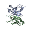
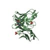
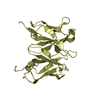
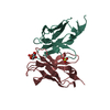
- PDB-1j05: The crystal structure of anti-carcinoembryonic antigen monoclonal... -
+ Open data

- Basic information
| Entry | Database: PDB / ID: 1j05 | ||||||
|---|---|---|---|---|---|---|---|
| Title | The crystal structure of anti-carcinoembryonic antigen monoclonal antibody T84.66 Fv fragment | ||||||
 Components Components |
| ||||||
 Keywords Keywords | IMMUNE SYSTEM / Immunoglobulin | ||||||
| Function / homology |  Function and homology information Function and homology informationimmunoglobulin complex / adaptive immune response / immune response / extracellular region / plasma membrane Similarity search - Function | ||||||
| Biological species |  | ||||||
| Method |  X-RAY DIFFRACTION / SYNCHROTRON / MOLECULAR REPLACEMENT / Resolution: 1.5 Å X-RAY DIFFRACTION / SYNCHROTRON / MOLECULAR REPLACEMENT / Resolution: 1.5 Å | ||||||
 Authors Authors | Kondo, H. / Nishimura, Y. / Shiroishi, M. / Asano, R. / Noro, N. / Tsumoto, K. / Kumagai, I. | ||||||
 Citation Citation | Journal: To be Published Title: The crystal structure of anti-carcinoembryonic antigen monoclonal antibody T84.66 Fv fragment Authors: Kondo, H. / Nishimura, Y. / Shiroishi, M. / Asano, R. / Noro, N. / Tsumoto, K. / Kumagai, I. | ||||||
| History |
|
- Structure visualization
| Structure viewer | Molecule: MolmilJmol/JSmol |
|---|
- Downloads & links
-Download
| PDBx/mmCIF format | 1j05.cif.gz | 106.6 KB | Display | PDBx/mmCIF format |
|---|---|---|---|---|
| PDB format | pdb1j05.ent.gz | 81.9 KB | Display | PDB format |
| PDBx/mmJSON format | 1j05.json.gz | Tree view | PDBx/mmJSON format | |
| Others |  Other downloads Other downloads |
-Validation report
| Arichive directory | https://data.pdbj.org/pub/pdb/validation_reports/j0/1j05ftp://data.pdbj.org/pub/pdb/validation_reports/j0/1j05 | HTTPS FTP |
|---|
-Related structure data
| Similar structure data |
|---|


-Links
 PDBj
PDBj

- Assembly
| Deposited unit | 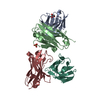
| ||||||||
|---|---|---|---|---|---|---|---|---|---|
| 1 | 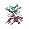
| ||||||||
| 2 | 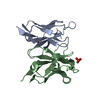
| ||||||||
| 3 | 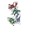
| ||||||||
| Unit cell |
| ||||||||
| Details | The biological assemblies are formed between chain L and H and between A and B. |
-Components
| #1: Antibody | Mass: 12113.562 Da / Num. of mol.: 2 / Fragment: Fv fragment Source method: isolated from a genetically manipulated source Source: (gene. exp.)  #2: Antibody | Mass: 13233.598 Da / Num. of mol.: 2 / Fragment: Fv fragment Source method: isolated from a genetically manipulated source Source: (gene. exp.) #3: Chemical |   Mass: 92.094 Da / Num. of mol.: 2 / Source method: obtained synthetically / Formula: C3H8O3 Mass: 92.094 Da / Num. of mol.: 2 / Source method: obtained synthetically / Formula: C3H8O3#4: Chemical |   Mass: 94.971 Da / Num. of mol.: 2 / Source method: obtained synthetically / Formula: PO4 Mass: 94.971 Da / Num. of mol.: 2 / Source method: obtained synthetically / Formula: PO4#5: Water | ChemComp-HOH / |  Mass: 18.015 Da / Num. of mol.: 256 / Source method: isolated from a natural source / Formula: H2O Mass: 18.015 Da / Num. of mol.: 256 / Source method: isolated from a natural source / Formula: H2OHas protein modification | Y | |
|---|
-Experimental details
-Experiment
| Experiment | Method: X-RAY DIFFRACTION / Number of used crystals: 1 |
|---|
- Sample preparation
| Crystal | Density Matthews: 2.7 Å3/Da / Density % sol: 54.04 % |
|---|---|
| Crystal grow | Temperature: 293 K / Method: vapor diffusion, hanging drop / pH: 7 Details: Na, K Phosphate, Na Hepes, Glycerol, pH 7.0, VAPOR DIFFUSION, HANGING DROP, temperature 293K |
-Data collection
| Diffraction | Mean temperature: 100 K |
|---|---|
| Diffraction source | Source: SYNCHROTRON / Site: Photon Factory  / Beamline: BL-6A / Wavelength: 1 Å / Beamline: BL-6A / Wavelength: 1 Å |
| Detector | Type: ADSC QUANTUM 4 / Detector: CCD / Date: Apr 23, 2002 |
| Radiation | Protocol: SINGLE WAVELENGTH / Monochromatic (M) / Laue (L): M / Scattering type: x-ray |
| Radiation wavelength | Wavelength: 1 Å / Relative weight: 1 |
| Reflection | Resolution: 1.5→23.25 Å / Num. all: 91647 / Num. obs: 91647 / % possible obs: 99.8 % / Observed criterion σ(F): 0 / Observed criterion σ(I): 0 / Redundancy: 3.6 % / Biso Wilson estimate: 16.7 Å2 / Rmerge(I) obs: 0.069 / Net I/σ(I): 6.7 |
| Reflection shell | Resolution: 1.5→1.58 Å / Redundancy: 3.5 % / Rmerge(I) obs: 0.325 / Mean I/σ(I) obs: 2.2 / Num. unique all: 13262 / % possible all: 99.8 |
- Processing
| Software |
| |||||||||||||||||||||
|---|---|---|---|---|---|---|---|---|---|---|---|---|---|---|---|---|---|---|---|---|---|---|
| Refinement | Method to determine structure: MOLECULAR REPLACEMENT / Resolution: 1.5→5 Å / σ(F): 2 / Stereochemistry target values: Engh & Huber
| |||||||||||||||||||||
| Refinement step | Cycle: LAST / Resolution: 1.5→5 Å
|
