 Movie
Movie Controller


+ Open data

- Basic information
| Entry | Database: PDB / ID: 6lfv | ||||||
|---|---|---|---|---|---|---|---|
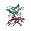
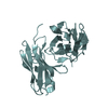
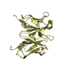
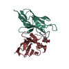
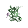
| Title | Crystal structure of PCB4scFv(hN56D) | ||||||
 Components Components | PCB4scFv(hN56D) | ||||||
 Keywords Keywords | IMMUNE SYSTEM / Co-PCB / antibody fragment | ||||||
| Function / homology | Immunoglobulins / Immunoglobulin-like / Sandwich / Mainly Beta Function and homology information Function and homology information | ||||||
| Biological species |  | ||||||
| Method |  X-RAY DIFFRACTION / SYNCHROTRON / MOLECULAR REPLACEMENT / Resolution: 1.9 Å X-RAY DIFFRACTION / SYNCHROTRON / MOLECULAR REPLACEMENT / Resolution: 1.9 Å | ||||||
 Authors Authors | Nakamura, T. / Yamagata, Y. / Morioka, H. | ||||||
| Funding support |  Japan, 1items Japan, 1items
| ||||||
 Citation Citation | Journal: To Be Published Title: Crystal structure of PCB4scFv(hN56D) Authors: Nakamura, T. / Morioka, H. | ||||||
| History |
|
- Structure visualization
| Structure viewer | Molecule: MolmilJmol/JSmol |
|---|
- Downloads & links
-Download
| PDBx/mmCIF format | 6lfv.cif.gz | 110.4 KB | Display | PDBx/mmCIF format |
|---|---|---|---|---|
| PDB format | pdb6lfv.ent.gz | 80 KB | Display | PDB format |
| PDBx/mmJSON format | 6lfv.json.gz | Tree view | PDBx/mmJSON format | |
| Others |  Other downloads Other downloads |
-Validation report
| Arichive directory | https://data.pdbj.org/pub/pdb/validation_reports/lf/6lfvftp://data.pdbj.org/pub/pdb/validation_reports/lf/6lfv | HTTPS FTP |
|---|
-Related structure data
| Related structure data | 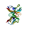 3esuS S: Starting model for refinement |
|---|---|
| Similar structure data |


-Links
 PDBj
PDBj

- Assembly
| Deposited unit | 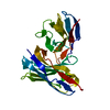
| ||||||||||||
|---|---|---|---|---|---|---|---|---|---|---|---|---|---|
| 1 |
| ||||||||||||
| Unit cell |
|
-Components
| #1: Antibody | Mass: 27237.428 Da / Num. of mol.: 1 Source method: isolated from a genetically manipulated source Source: (gene. exp.)  |
|---|---|
| #2: Water | ChemComp-HOH /  Mass: 18.015 Da / Num. of mol.: 107 / Source method: isolated from a natural source / Formula: H2O Mass: 18.015 Da / Num. of mol.: 107 / Source method: isolated from a natural source / Formula: H2O |
| Has protein modification | Y |
-Experimental details
-Experiment
| Experiment | Method: X-RAY DIFFRACTION / Number of used crystals: 1 |
|---|
- Sample preparation
| Crystal | Density Matthews: 2.11 Å3/Da / Density % sol: 41.71 % |
|---|---|
| Crystal grow | Temperature: 293 K / Method: vapor diffusion, hanging drop / Details: HEPES, PEG 8000, ethylene glycol |
-Data collection
| Diffraction | Mean temperature: 100 K / Serial crystal experiment: N |
|---|---|
| Diffraction source | Source: SYNCHROTRON / Site: Photon Factory / Beamline: BL-5A / Wavelength: 1 Å |
| Detector | Type: ADSC QUANTUM 315 / Detector: CCD / Date: Feb 26, 2016 |
| Radiation | Protocol: SINGLE WAVELENGTH / Monochromatic (M) / Laue (L): M / Scattering type: x-ray |
| Radiation wavelength | Wavelength: 1 Å / Relative weight: 1 |
| Reflection | Resolution: 1.9→32.78 Å / Num. obs: 41553 / % possible obs: 99.9 % / Redundancy: 7.4 % / Biso Wilson estimate: 29.21 Å2 / Rmerge(I) obs: 0.048 / Net I/σ(I): 25.8 |
| Reflection shell | Resolution: 1.9→1.95 Å / Rmerge(I) obs: 0.527 / Num. unique obs: 1322 |
- Processing
| Software |
| ||||||||||||||||||||||||
|---|---|---|---|---|---|---|---|---|---|---|---|---|---|---|---|---|---|---|---|---|---|---|---|---|---|
| Refinement | Method to determine structure: MOLECULAR REPLACEMENT Starting model: 3ESU Resolution: 1.9→32.78 Å / SU ML: 0.2093 / Cross valid method: FREE R-VALUE / σ(F): 1.22 / Phase error: 23.7117
| ||||||||||||||||||||||||
| Solvent computation | Shrinkage radii: 0.9 Å / VDW probe radii: 1.11 Å | ||||||||||||||||||||||||
| Displacement parameters | Biso mean: 42.15 Å2 | ||||||||||||||||||||||||
| Refinement step | Cycle: LAST / Resolution: 1.9→32.78 Å
| ||||||||||||||||||||||||
| Refine LS restraints |
| ||||||||||||||||||||||||
| LS refinement shell | Resolution: 1.9→1.95 Å
|
