 Movie
Movie Controller

[English] 日本語
 Yorodumi
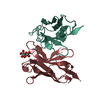
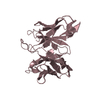
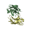
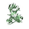
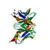
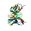
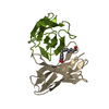
- PDB-3esu: Crystal structure of anthrax-neutralizing single-chain antibody 14b7 -
+ Open data

- Basic information
| Entry | Database: PDB / ID: 3esu | ||||||||||||||||||||
|---|---|---|---|---|---|---|---|---|---|---|---|---|---|---|---|---|---|---|---|---|---|
| Title | Crystal structure of anthrax-neutralizing single-chain antibody 14b7 | ||||||||||||||||||||
 Components Components | Antibody 14b7* Keywords KeywordsIMMUNE SYSTEM / single-chain FV / monoclonal antibody / immunoglobulin | Function / homology | Immunoglobulins / Immunoglobulin-like / Sandwich / Mainly Beta |  Function and homology information Function and homology informationBiological species |  Method |  X-RAY DIFFRACTION / SYNCHROTRON / MOLECULAR REPLACEMENT / Resolution: 1.3 Å X-RAY DIFFRACTION / SYNCHROTRON / MOLECULAR REPLACEMENT / Resolution: 1.3 Å  Authors AuthorsMonzingo, A.F. / Maynard, J.A. / Iverson, B.L. / Georgiou, G. / Robertus, J.D. |  CitationJournal: J.Mol.Biol. / Year: 2009 CitationJournal: J.Mol.Biol. / Year: 2009Title: Crystal structure of the engineered neutralizing antibody m18 complexed to domain 4 of the anthrax protective antigen. Authors: Leysath, C.E. / Monzingo, A.F. / Maynard, J.A. / Barnett, J. / Georgiou, G. / Iverson, B.L. / Robertus, J.D. History |
|
- Structure visualization
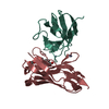
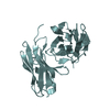
| Structure viewer | Molecule: MolmilJmol/JSmol |
|---|
- Downloads & links
-Download
| PDBx/mmCIF format | 3esu.cif.gz | 62 KB | Display | PDBx/mmCIF format |
|---|---|---|---|---|
| PDB format | pdb3esu.ent.gz | 43.9 KB | Display | PDB format |
| PDBx/mmJSON format | 3esu.json.gz | Tree view | PDBx/mmJSON format | |
| Others |  Other downloads Other downloads |
-Validation report
| Arichive directory | https://data.pdbj.org/pub/pdb/validation_reports/es/3esuftp://data.pdbj.org/pub/pdb/validation_reports/es/3esu | HTTPS FTP |
|---|
-Related structure data
| Related structure data |  3esvC  3et9C  3etbC  1forS  1jv5S C: citing same article ( S: Starting model for refinement |
|---|---|
| Similar structure data |

-Links
 PDBj
PDBj

- Assembly
| Deposited unit | 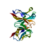
| ||||||||
|---|---|---|---|---|---|---|---|---|---|
| 1 |
| ||||||||
| Unit cell |
| ||||||||
| Details | Asymmetric unit contains one biological unit |
-Components
| #1: Antibody | Mass: 26561.275 Da / Num. of mol.: 1 / Mutation: Q3V, M4L, T5I, Q78L, K107R, L1045P Source method: isolated from a genetically manipulated source Details: periplasm / Source: (gene. exp.)  |
|---|---|
| #2: Water | ChemComp-HOH /  Mass: 18.015 Da / Num. of mol.: 265 / Source method: isolated from a natural source / Formula: H2O Mass: 18.015 Da / Num. of mol.: 265 / Source method: isolated from a natural source / Formula: H2O |
| Has protein modification | Y |
| Sequence details | RESIDUE NUMBERS FOR ANTIBODY 14B7* HEAVY CHAIN FRAGMENT OF ENTITY 1 HAVE OFFSET 1000 TO DISTINGUISH ...RESIDUE NUMBERS FOR ANTIBODY 14B7* HEAVY CHAIN FRAGMENT OF ENTITY 1 HAVE OFFSET 1000 TO DISTINGUIS |
-Experimental details
-Experiment
| Experiment | Method: X-RAY DIFFRACTION / Number of used crystals: 1 |
|---|
- Sample preparation
| Crystal | Density Matthews: 2.05 Å3/Da / Density % sol: 40.05 % |
|---|---|
| Crystal grow | Temperature: 298 K / Method: vapor diffusion, sitting drop / pH: 7.5 Details: 20% PEG 4000, 0.1 M HEPES, 10% Isopropanol, pH 7.5, VAPOR DIFFUSION, SITTING DROP, temperature 298K |
-Data collection
| Diffraction | Mean temperature: 100 K |
|---|---|
| Diffraction source | Source: SYNCHROTRON / Site: ALS  / Beamline: 8.2.1 / Wavelength: 0.9641 Å / Beamline: 8.2.1 / Wavelength: 0.9641 Å |
| Detector | Type: ADSC QUANTUM 210 / Detector: CCD / Date: Dec 14, 2003 |
| Radiation | Protocol: SINGLE WAVELENGTH / Monochromatic (M) / Laue (L): M / Scattering type: x-ray |
| Radiation wavelength | Wavelength: 0.9641 Å / Relative weight: 1 |
| Reflection | Resolution: 1.3→50 Å / Num. obs: 54301 / % possible obs: 99.1 % / Observed criterion σ(I): 0 / Redundancy: 12.8 % / Rmerge(I) obs: 0.079 / Χ2: 1.455 / Net I/σ(I): 9.4 |
| Reflection shell | Resolution: 1.3→1.35 Å / Rmerge(I) obs: 0.466 / Mean I/σ(I) obs: 4.5 / Num. unique all: 5384 / Χ2: 0.825 / % possible all: 100 |
- Processing
| Software |
| ||||||||||||||||||||||||||||
|---|---|---|---|---|---|---|---|---|---|---|---|---|---|---|---|---|---|---|---|---|---|---|---|---|---|---|---|---|---|
| Refinement | Method to determine structure: MOLECULAR REPLACEMENT Starting model: PDB entries 1JV5, 1FOR Resolution: 1.3→20 Å / Occupancy max: 1 / Occupancy min: 1 / FOM work R set: 0.86 / Isotropic thermal model: isotropic / Cross valid method: THROUGHOUT / σ(F): 0 / Stereochemistry target values: Engh & Huber
| ||||||||||||||||||||||||||||
| Solvent computation | Bsol: 36.343 Å2 | ||||||||||||||||||||||||||||
| Displacement parameters | Biso max: 24.24 Å2 / Biso mean: 11.318 Å2 / Biso min: 4.95 Å2
| ||||||||||||||||||||||||||||
| Refinement step | Cycle: LAST / Resolution: 1.3→20 Å
| ||||||||||||||||||||||||||||
| Refine LS restraints |
| ||||||||||||||||||||||||||||
| Xplor file |
|
