 Movie
Movie Controller


+ Open data

- Basic information
| Entry | Database: PDB / ID: 2bfv | ||||||
|---|---|---|---|---|---|---|---|
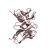
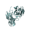
| Title | MONOCLONAL ANTIBODY FRAGMENT FV4155 FROM E. COLI | ||||||
 Components Components | (FV4155) x 2 | ||||||
 Keywords Keywords | IMMUNOGLOBULIN / FV FRAGMENT / STEROID HORMONE / FINE SPECIFICITY | ||||||
| Function / homology |  Function and homology information Function and homology informationimmunoglobulin complex / antigen binding / adaptive immune response / immune response / extracellular region / plasma membrane Similarity search - Function | ||||||
| Biological species |  | ||||||
| Method |  X-RAY DIFFRACTION / MOLECULAR REPLACEMENT / Resolution: 2.5 Å X-RAY DIFFRACTION / MOLECULAR REPLACEMENT / Resolution: 2.5 Å | ||||||
 Authors Authors | Trinh, C.H. / Phillips, S.E.V. | ||||||
 Citation Citation | Journal: Structure / Year: 1997 Title: Antibody fragment Fv4155 bound to two closely related steroid hormones: the structural basis of fine specificity. Authors: Trinh, C.H. / Hemmington, S.D. / Verhoeyen, M.E. / Phillips, S.E. | ||||||
| History |
|
- Structure visualization
| Structure viewer | Molecule: MolmilJmol/JSmol |
|---|
- Downloads & links
-Download
| PDBx/mmCIF format | 2bfv.cif.gz | 60.4 KB | Display | PDBx/mmCIF format |
|---|---|---|---|---|
| PDB format | pdb2bfv.ent.gz | 43.4 KB | Display | PDB format |
| PDBx/mmJSON format | 2bfv.json.gz | Tree view | PDBx/mmJSON format | |
| Others |  Other downloads Other downloads |
-Validation report
| Arichive directory | https://data.pdbj.org/pub/pdb/validation_reports/bf/2bfvftp://data.pdbj.org/pub/pdb/validation_reports/bf/2bfv | HTTPS FTP |
|---|
-Related structure data
| Related structure data |  1bfvC 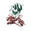 1cfvC 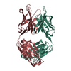 1nbvS C: citing same article ( S: Starting model for refinement |
|---|---|
| Similar structure data |


-Links
 PDBj
PDBj

- Assembly
| Deposited unit | 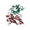
| ||||||||
|---|---|---|---|---|---|---|---|---|---|
| 1 |
| ||||||||
| Unit cell |
|
-Components
| #1: Antibody | Mass: 12437.998 Da / Num. of mol.: 1 / Fragment: MONOCLONAL ANTIBODY FV FRAGMENT Source method: isolated from a genetically manipulated source Source: (gene. exp.)  |
|---|---|
| #2: Antibody | Mass: 13191.760 Da / Num. of mol.: 1 / Fragment: MONOCLONAL ANTIBODY FV FRAGMENT Source method: isolated from a genetically manipulated source Source: (gene. exp.) |
| #3: Chemical | ChemComp-STG /   Mass: 464.505 Da / Num. of mol.: 1 / Source method: obtained synthetically / Formula: C24H32O9 Mass: 464.505 Da / Num. of mol.: 1 / Source method: obtained synthetically / Formula: C24H32O9 |
| #4: Water | ChemComp-HOH /  Mass: 18.015 Da / Num. of mol.: 55 / Source method: isolated from a natural source / Formula: H2O Mass: 18.015 Da / Num. of mol.: 55 / Source method: isolated from a natural source / Formula: H2O |
| Has protein modification | Y |
-Experimental details
-Experiment
| Experiment | Method: X-RAY DIFFRACTION / Number of used crystals: 1 |
|---|
- Sample preparation
| Crystal | Density Matthews: 3.14 Å3/Da / Density % sol: 60.8 % | ||||||||||||||||||||||||||||||||||||||||
|---|---|---|---|---|---|---|---|---|---|---|---|---|---|---|---|---|---|---|---|---|---|---|---|---|---|---|---|---|---|---|---|---|---|---|---|---|---|---|---|---|---|
| Crystal grow | pH: 8.5 Details: PROTEIN WAS CRYSTALLIZED FROM 15% (W/V) PEG 4000, 100MM NA ACETATE AND 50MM TRIS HCL PH 8.5 | ||||||||||||||||||||||||||||||||||||||||
| Crystal grow | *PLUS Method: vapor diffusion, hanging drop | ||||||||||||||||||||||||||||||||||||||||
| Components of the solutions | *PLUS
|
-Data collection
| Diffraction | Mean temperature: 293 K |
|---|---|
| Diffraction source | Source: ROTATING ANODE / Type: RIGAKU RUH2R / Wavelength: 1.5418 |
| Detector | Type: RIGAKU / Detector: IMAGE PLATE / Date: Apr 1, 1995 / Details: MSC DOUBLE FOCUSING MIRRORS |
| Radiation | Monochromator: NI FILTER / Monochromatic (M) / Laue (L): M / Scattering type: x-ray |
| Radiation wavelength | Wavelength: 1.5418 Å / Relative weight: 1 |
| Reflection | Resolution: 2.5→20 Å / Num. obs: 7836 / % possible obs: 86.9 % / Observed criterion σ(I): 3 / Redundancy: 1.5 % / Rsym value: 0.064 / Net I/σ(I): 7.21 |
| Reflection shell | Resolution: 2.5→2.59 Å / Redundancy: 1.5 % / Mean I/σ(I) obs: 2.6 / Rsym value: 0.225 / % possible all: 87.5 |
| Reflection | *PLUS Num. measured all: 12233 / Rmerge(I) obs: 0.064 |
| Reflection shell | *PLUS % possible obs: 87.5 % / Rmerge(I) obs: 0.225 |
- Processing
| Software |
| ||||||||||||||||||||||||||||||||||||||||||||||||||||||||||||||||||||||||||||||||||||
|---|---|---|---|---|---|---|---|---|---|---|---|---|---|---|---|---|---|---|---|---|---|---|---|---|---|---|---|---|---|---|---|---|---|---|---|---|---|---|---|---|---|---|---|---|---|---|---|---|---|---|---|---|---|---|---|---|---|---|---|---|---|---|---|---|---|---|---|---|---|---|---|---|---|---|---|---|---|---|---|---|---|---|---|---|---|
| Refinement | Method to determine structure: MOLECULAR REPLACEMENT Starting model: PDB ENTRY 1NBV Resolution: 2.5→10 Å / σ(F): 0 Details: VAL L 56 HAS DIHEDRAL ANGLES WHICH LIE OUTSIDE THEIR EXPECTED RANGE IN THE GAMMA QUADRANT OF THE RAMACHANDRAN PLOT.
| ||||||||||||||||||||||||||||||||||||||||||||||||||||||||||||||||||||||||||||||||||||
| Displacement parameters | Biso mean: 27.44 Å2 | ||||||||||||||||||||||||||||||||||||||||||||||||||||||||||||||||||||||||||||||||||||
| Refinement step | Cycle: LAST / Resolution: 2.5→10 Å
| ||||||||||||||||||||||||||||||||||||||||||||||||||||||||||||||||||||||||||||||||||||
| Refine LS restraints |
| ||||||||||||||||||||||||||||||||||||||||||||||||||||||||||||||||||||||||||||||||||||
| Software | *PLUS Name: PROLSQ(CCP4) / Classification: refinement | ||||||||||||||||||||||||||||||||||||||||||||||||||||||||||||||||||||||||||||||||||||
| Refinement | *PLUS Rfactor obs: 0.176 | ||||||||||||||||||||||||||||||||||||||||||||||||||||||||||||||||||||||||||||||||||||
| Solvent computation | *PLUS | ||||||||||||||||||||||||||||||||||||||||||||||||||||||||||||||||||||||||||||||||||||
| Displacement parameters | *PLUS |
