 Movie
Movie Controller

[English] 日本語
 Yorodumi
- PDB-6ffj: Anti-tumor antibody 14F7-derived single chain fragment variable (scFv) -
+ Open data

- Basic information
| Entry | Database: PDB / ID: 6ffj | ||||||
|---|---|---|---|---|---|---|---|
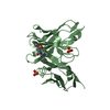
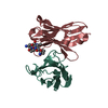
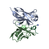
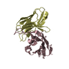
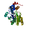
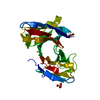
| Title | Anti-tumor antibody 14F7-derived single chain fragment variable (scFv) | ||||||
 Components Components | 14F7-derived scFv | ||||||
 Keywords Keywords | ANTITUMOR PROTEIN / ganglioside / scfv / antibody fragment / single chain fragment variable / immuno therapy / 14F7 / gm3 | ||||||
| Function / homology | Immunoglobulins / Immunoglobulin-like / Sandwich / Mainly Beta Function and homology information Function and homology information | ||||||
| Biological species |  Homo sapiens (human) Homo sapiens (human) | ||||||
| Method |  X-RAY DIFFRACTION / SYNCHROTRON / MOLECULAR REPLACEMENT / Resolution: 2.2 Å X-RAY DIFFRACTION / SYNCHROTRON / MOLECULAR REPLACEMENT / Resolution: 2.2 Å | ||||||
 Authors Authors | Bjerregaard-Andersen, K. / Heggelund, J.E. / Krengel, U. | ||||||
 Citation Citation | Journal: Sci Rep / Year: 2018 Title: Crystal structure of an L chain optimised 14F7 anti-ganglioside Fv suggests a unique tumour-specificity through an unusual H-chain CDR3 architecture. Authors: Bjerregaard-Andersen, K. / Johannesen, H. / Abdel-Rahman, N. / Heggelund, J.E. / Hoas, H.M. / Abraha, F. / Bousquet, P.A. / Hoydahl, L.S. / Burschowsky, D. / Rojas, G. / Oscarson, S. / Loset, G.A. / Krengel, U. | ||||||
| History |
|
- Structure visualization
| Structure viewer | Molecule: MolmilJmol/JSmol |
|---|
- Downloads & links
-Download
| PDBx/mmCIF format | 6ffj.cif.gz | 372.8 KB | Display | PDBx/mmCIF format |
|---|---|---|---|---|
| PDB format | pdb6ffj.ent.gz | 304.9 KB | Display | PDB format |
| PDBx/mmJSON format | 6ffj.json.gz | Tree view | PDBx/mmJSON format | |
| Others |  Other downloads Other downloads |
-Validation report
| Arichive directory | https://data.pdbj.org/pub/pdb/validation_reports/ff/6ffjftp://data.pdbj.org/pub/pdb/validation_reports/ff/6ffj | HTTPS FTP |
|---|
-Related structure data
| Related structure data |  3umtS S: Starting model for refinement |
|---|---|
| Similar structure data |


-Links
 PDBj
PDBj

- Assembly
| Deposited unit | 
| ||||||||
|---|---|---|---|---|---|---|---|---|---|
| 1 | 
| ||||||||
| 2 | 
| ||||||||
| 3 | 
| ||||||||
| 4 | 
| ||||||||
| Unit cell |
|
-Components
| #1: Antibody | Mass: 28116.238 Da / Num. of mol.: 4 Source method: isolated from a genetically manipulated source Details: Humanized antibody fragment / Source: (gene. exp.) Homo sapiens (human) / Plasmid: pFKPENDetails (production host): co-expresses E. coli chaperone FkpA Production host:  #2: Water | ChemComp-HOH / |  Mass: 18.015 Da / Num. of mol.: 254 / Source method: isolated from a natural source / Formula: H2O Mass: 18.015 Da / Num. of mol.: 254 / Source method: isolated from a natural source / Formula: H2OHas protein modification | Y | |
|---|
-Experimental details
-Experiment
| Experiment | Method: X-RAY DIFFRACTION / Number of used crystals: 1 |
|---|
- Sample preparation
| Crystal | Density Matthews: 2.53 Å3/Da / Density % sol: 51.37 % / Description: Plates |
|---|---|
| Crystal grow | Temperature: 293 K / Method: vapor diffusion, sitting drop Details: 12.5 % w/v PEG 1000, 12.5 % w/v PEG 3350, 12.5 % v/v MPD, 0.02 M 1,6-hexandiol, 0.02 M 1-butanol, 0.02 M (RS)-1,2-propanediol, 0.02 M 2-propanol, 0.02 M 1,4-butanediol, 0.02 M 1,3- ...Details: 12.5 % w/v PEG 1000, 12.5 % w/v PEG 3350, 12.5 % v/v MPD, 0.02 M 1,6-hexandiol, 0.02 M 1-butanol, 0.02 M (RS)-1,2-propanediol, 0.02 M 2-propanol, 0.02 M 1,4-butanediol, 0.02 M 1,3-propanediol, 0.1 M bicine/tris base pH 8.5 |
-Data collection
| Diffraction | Mean temperature: 100 K |
|---|---|
| Diffraction source | Source: SYNCHROTRON / Site: ESRF  / Beamline: ID29 / Wavelength: 1.0332 Å / Beamline: ID29 / Wavelength: 1.0332 Å |
| Detector | Type: DECTRIS PILATUS3 S 6M / Detector: PIXEL / Date: Feb 5, 2017 |
| Radiation | Protocol: SINGLE WAVELENGTH / Monochromatic (M) / Laue (L): M / Scattering type: x-ray |
| Radiation wavelength | Wavelength: 1.0332 Å / Relative weight: 1 |
| Reflection | Resolution: 2.2→46.71 Å / Num. obs: 49026 / % possible obs: 94.8 % / Redundancy: 2.5 % / Biso Wilson estimate: 37.78 Å2 / CC1/2: 0.989 / Rmerge(I) obs: 0.112 / Rpim(I) all: 0.078 / Rrim(I) all: 0.137 / Net I/σ(I): 3.4 |
| Reflection shell | Resolution: 2.2→2.28 Å / Redundancy: 2.5 % / Rmerge(I) obs: 0.532 / Mean I/σ(I) obs: 1.2 / Num. unique obs: 4880 / CC1/2: 0.8 / Rpim(I) all: 0.374 / Rrim(I) all: 0.653 / % possible all: 96.4 |
- Processing
| Software |
| |||||||||||||||||||||||||||||||||||||||||||||||||||||||||||||||||||||||||||||||||||||||||||||||||||||||||||||||||||||||||||||||||||||
|---|---|---|---|---|---|---|---|---|---|---|---|---|---|---|---|---|---|---|---|---|---|---|---|---|---|---|---|---|---|---|---|---|---|---|---|---|---|---|---|---|---|---|---|---|---|---|---|---|---|---|---|---|---|---|---|---|---|---|---|---|---|---|---|---|---|---|---|---|---|---|---|---|---|---|---|---|---|---|---|---|---|---|---|---|---|---|---|---|---|---|---|---|---|---|---|---|---|---|---|---|---|---|---|---|---|---|---|---|---|---|---|---|---|---|---|---|---|---|---|---|---|---|---|---|---|---|---|---|---|---|---|---|---|---|
| Refinement | Method to determine structure: MOLECULAR REPLACEMENT Starting model: 3UMT Resolution: 2.2→46.708 Å / SU ML: 0.28 / Cross valid method: FREE R-VALUE / σ(F): 1.33 / Phase error: 28.59 / Stereochemistry target values: ML
| |||||||||||||||||||||||||||||||||||||||||||||||||||||||||||||||||||||||||||||||||||||||||||||||||||||||||||||||||||||||||||||||||||||
| Solvent computation | Shrinkage radii: 0.9 Å / VDW probe radii: 1.11 Å / Solvent model: FLAT BULK SOLVENT MODEL | |||||||||||||||||||||||||||||||||||||||||||||||||||||||||||||||||||||||||||||||||||||||||||||||||||||||||||||||||||||||||||||||||||||
| Refinement step | Cycle: LAST / Resolution: 2.2→46.708 Å
| |||||||||||||||||||||||||||||||||||||||||||||||||||||||||||||||||||||||||||||||||||||||||||||||||||||||||||||||||||||||||||||||||||||
| Refine LS restraints |
| |||||||||||||||||||||||||||||||||||||||||||||||||||||||||||||||||||||||||||||||||||||||||||||||||||||||||||||||||||||||||||||||||||||
| LS refinement shell |
|
