 Movie
Movie Controller

[English] 日本語
 Yorodumi
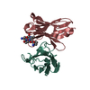
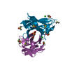
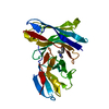
- PDB-4r3s: Crystal Structure of anti-MSP2 Fv fragment (mAb6D8)in complex wit... -
+ Open data

- Basic information
| Entry | Database: PDB / ID: 4r3s | ||||||
|---|---|---|---|---|---|---|---|
| Title | Crystal Structure of anti-MSP2 Fv fragment (mAb6D8)in complex with MSP2 11-23 | ||||||
 Components Components |
| ||||||
 Keywords Keywords | IMMUNE SYSTEM / IMMUNOGLOBULIN FOLD / N-TERMINAL MSP2 / UNSTRUCTURED ANTIGEN | ||||||
| Function / homology |  Function and homology information Function and homology information | ||||||
| Biological species |   | ||||||
| Method |  X-RAY DIFFRACTION / SYNCHROTRON / MOLECULAR REPLACEMENT / Resolution: 1.7 Å X-RAY DIFFRACTION / SYNCHROTRON / MOLECULAR REPLACEMENT / Resolution: 1.7 Å | ||||||
 Authors Authors | Morales, R.A.V. / MacRaild, C.A. / Seow, J. / Bankala, K. / Drinkwater, N. / McGowan, S. / Rouet, R. / Christ, D. / Anders, R.F. / Norton, R.S. | ||||||
 Citation Citation | Journal: Sci Rep / Year: 2015 Title: Structural basis for epitope masking and strain specificity of a conserved epitope in an intrinsically disordered malaria vaccine candidate Authors: Morales, R.A.V. / MacRaild, C.A. / Seow, J. / Krishnarjuna, B. / Drinkwater, N. / Rouet, R. / Anders, R.F. / Christ, D. / McGowan, S. / Norton, R.S. | ||||||
| History |
|
- Structure visualization
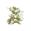
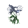
| Structure viewer | Molecule: MolmilJmol/JSmol |
|---|
- Downloads & links
-Download
| PDBx/mmCIF format | 4r3s.cif.gz | 119.1 KB | Display | PDBx/mmCIF format |
|---|---|---|---|---|
| PDB format | pdb4r3s.ent.gz | 91.6 KB | Display | PDB format |
| PDBx/mmJSON format | 4r3s.json.gz | Tree view | PDBx/mmJSON format | |
| Others |  Other downloads Other downloads |
-Validation report
| Arichive directory | https://data.pdbj.org/pub/pdb/validation_reports/r3/4r3sftp://data.pdbj.org/pub/pdb/validation_reports/r3/4r3s | HTTPS FTP |
|---|
-Related structure data
| Related structure data |  4qxtC  4qy8C  4qyoSC C: citing same article ( S: Starting model for refinement |
|---|---|
| Similar structure data |


-Links
 PDBj
PDBj

- Assembly
| Deposited unit | 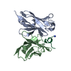
| ||||||||
|---|---|---|---|---|---|---|---|---|---|
| 1 |
| ||||||||
| Unit cell |
|
-Components
| #1: Antibody | Mass: 12489.899 Da / Num. of mol.: 1 Source method: isolated from a genetically manipulated source Source: (gene. exp.)  |
|---|---|
| #2: Antibody | Mass: 11959.071 Da / Num. of mol.: 1 Source method: isolated from a genetically manipulated source Source: (gene. exp.) |
| #3: Protein/peptide | Mass: 1611.849 Da / Num. of mol.: 1 / Fragment: 11-23 / Source method: obtained synthetically Details: This sequence occurs naturally in Plasmodium Falciparum Source: (synth.) References: UniProt: Q9GQZ3 |
| #4: Water | ChemComp-HOH /  Mass: 18.015 Da / Num. of mol.: 370 / Source method: isolated from a natural source / Formula: H2O Mass: 18.015 Da / Num. of mol.: 370 / Source method: isolated from a natural source / Formula: H2O |
| Has protein modification | Y |
| Sequence details | IN THIS PROTEIN, THE SEQUENCES OF THE ENTITY1 AND ENTITY2 WERE NOT AVAILABLE AT THE UNIPROT ...IN THIS PROTEIN, THE SEQUENCES OF THE ENTITY1 AND ENTITY2 WERE NOT AVAILABLE AT THE UNIPROT KNOWLEDGEB |
-Experimental details
-Experiment
| Experiment | Method: X-RAY DIFFRACTION / Number of used crystals: 1 |
|---|
- Sample preparation
| Crystal | Density Matthews: 1.95 Å3/Da / Density % sol: 37.02 % |
|---|---|
| Crystal grow | Temperature: 293 K / Method: vapor diffusion, hanging drop / pH: 4 Details: 30% PEG 8000, 0.2M NACL, 0.1M NAOAC, pH 4.0, VAPOR DIFFUSION, HANGING DROP, temperature 293K |
-Data collection
| Diffraction | Mean temperature: 100 K |
|---|---|
| Diffraction source | Source: SYNCHROTRON / Site: Australian Synchrotron  / Beamline: MX1 / Wavelength: 0.9537 Å / Beamline: MX1 / Wavelength: 0.9537 Å |
| Detector | Type: ADSC QUANTUM 210r / Detector: CCD / Date: May 9, 2014 |
| Radiation | Protocol: SINGLE WAVELENGTH / Monochromatic (M) / Laue (L): M / Scattering type: x-ray |
| Radiation wavelength | Wavelength: 0.9537 Å / Relative weight: 1 |
| Reflection | Resolution: 1.7→52.315 Å / Num. all: 23184 / Num. obs: 23184 / % possible obs: 100 % / Observed criterion σ(F): 1.35 / Redundancy: 33.8 % / Rmerge(I) obs: 0.8825 |
| Reflection shell | Resolution: 1.7→1.761 Å / Redundancy: 32.4 % / Rmerge(I) obs: 2.01 / Mean I/σ(I) obs: 5.15 / Num. unique all: 2279 / % possible all: 100 |
- Processing
| Software |
| ||||||||||||||||||||||||||||||||||||||||||||||||||
|---|---|---|---|---|---|---|---|---|---|---|---|---|---|---|---|---|---|---|---|---|---|---|---|---|---|---|---|---|---|---|---|---|---|---|---|---|---|---|---|---|---|---|---|---|---|---|---|---|---|---|---|
| Refinement | Method to determine structure: MOLECULAR REPLACEMENT Starting model: 4QYO Resolution: 1.7→52.315 Å / SU ML: 0.17 / σ(F): 1.35 / Phase error: 17.57 / Stereochemistry target values: Engh & Huber
| ||||||||||||||||||||||||||||||||||||||||||||||||||
| Solvent computation | Shrinkage radii: 0.9 Å / VDW probe radii: 1.11 Å / Solvent model: FLAT BULK SOLVENT MODEL | ||||||||||||||||||||||||||||||||||||||||||||||||||
| Refinement step | Cycle: LAST / Resolution: 1.7→52.315 Å
| ||||||||||||||||||||||||||||||||||||||||||||||||||
| Refine LS restraints |
| ||||||||||||||||||||||||||||||||||||||||||||||||||
| LS refinement shell | Refine-ID: X-RAY DIFFRACTION / Total num. of bins used: 9 / % reflection obs: 100 %
| ||||||||||||||||||||||||||||||||||||||||||||||||||
| Refinement TLS params. | Method: refined / Origin x: -4.4092 Å / Origin y: -1.4233 Å / Origin z: 13.1768 Å
| ||||||||||||||||||||||||||||||||||||||||||||||||||
| Refinement TLS group | Selection details: all |
