 Movie
Movie Controller

[English] 日本語
 Yorodumi
- PDB-2qst: Crystal structure of the V39C mutant of the N-terminal domain of ... -
+ Open data

- Basic information
| Entry | Database: PDB / ID: 2qst | ||||||
|---|---|---|---|---|---|---|---|
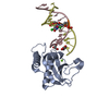
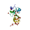
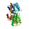
| Title | Crystal structure of the V39C mutant of the N-terminal domain of carcinoembryonic antigen (CEA) | ||||||
 Components Components | Carcinoembryonic antigen-related cell adhesion molecule 5 | ||||||
 Keywords Keywords | CELL ADHESION / Glycoprotein / GPI-anchor / Immunoglobulin domain / Lipoprotein / Membrane / Polymorphism | ||||||
| Function / homology |  Function and homology information Function and homology informationGPI anchor binding / homotypic cell-cell adhesion / negative regulation of myotube differentiation / Post-translational modification: synthesis of GPI-anchored proteins / heterophilic cell-cell adhesion / homophilic cell-cell adhesion / negative regulation of anoikis / side of membrane / Cell surface interactions at the vascular wall / basolateral plasma membrane ...GPI anchor binding / homotypic cell-cell adhesion / negative regulation of myotube differentiation / Post-translational modification: synthesis of GPI-anchored proteins / heterophilic cell-cell adhesion / homophilic cell-cell adhesion / negative regulation of anoikis / side of membrane / Cell surface interactions at the vascular wall / basolateral plasma membrane / apical plasma membrane / apoptotic process / negative regulation of apoptotic process / cell surface / protein homodimerization activity / extracellular exosome / extracellular region / membrane / identical protein binding / plasma membrane Similarity search - Function | ||||||
| Biological species |  Homo sapiens (human) Homo sapiens (human) | ||||||
| Method |  X-RAY DIFFRACTION / Resolution: 2.9 Å X-RAY DIFFRACTION / Resolution: 2.9 Å | ||||||
 Authors Authors | Le Trong, I. / Korotkova, N. / Moseley, S.L. / Stenkamp, R.E. | ||||||
 Citation Citation | Journal: Mol.Microbiol. / Year: 2008 Title: Binding of Dr adhesins of Escherichia coli to carcinoembryonic antigen triggers receptor dissociation. Authors: Korotkova, N. / Yang, Y. / Le Trong, I. / Cota, E. / Demeler, B. / Marchant, J. / Thomas, W.E. / Stenkamp, R.E. / Moseley, S.L. / Matthews, S. | ||||||
| History |
| ||||||
| Remark 999 | Sequence The sequence was obtained from human cDNA and the authors confirmed that ALA is the ...Sequence The sequence was obtained from human cDNA and the authors confirmed that ALA is the correct sequence. However, the authors do not know if it is a mutation introduced by PCR or whether it is an SNP that results in the different amino acid. |
- Structure visualization
| Structure viewer | Molecule: MolmilJmol/JSmol |
|---|
- Downloads & links
-Download
| PDBx/mmCIF format | 2qst.cif.gz | 56.1 KB | Display | PDBx/mmCIF format |
|---|---|---|---|---|
| PDB format | pdb2qst.ent.gz | 42.7 KB | Display | PDB format |
| PDBx/mmJSON format | 2qst.json.gz | Tree view | PDBx/mmJSON format | |
| Others |  Other downloads Other downloads |
-Validation report
| Arichive directory | https://data.pdbj.org/pub/pdb/validation_reports/qs/2qstftp://data.pdbj.org/pub/pdb/validation_reports/qs/2qst | HTTPS FTP |
|---|
-Related structure data


-Links
 PDBj
PDBj


- Assembly
| Deposited unit | 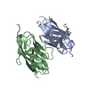
| ||||||||
|---|---|---|---|---|---|---|---|---|---|
| 1 | 
| ||||||||
| 2 | 
| ||||||||
| Unit cell |
| ||||||||
| Details | The two chains in the asymmetric unit (A,B) are parts of two biological units. A (x,y,z) and its crystallographically related partner, A'(x-y,-y,-z) form one dimer. B (x,y,z) and its crystallographically related partner, B' (2/3-x,1/3+y-x,1/3-z) form a similar, but slightly different, dimer. |
-Components
| #1: Protein | Mass: 12435.001 Da / Num. of mol.: 2 / Fragment: N-terminal domain of CEA (UNP residues 34-110) / Mutation: V39C Source method: isolated from a genetically manipulated source Source: (gene. exp.) Homo sapiens (human) / Gene: CEACAM5, CEA / Plasmid: pET-21d / Species (production host): Escherichia coli / Production host:  #2: Water | ChemComp-HOH / |  Mass: 18.015 Da / Num. of mol.: 11 / Source method: isolated from a natural source / Formula: H2O Mass: 18.015 Da / Num. of mol.: 11 / Source method: isolated from a natural source / Formula: H2O |
|---|
-Experimental details
-Experiment
| Experiment | Method: X-RAY DIFFRACTION / Number of used crystals: 1 |
|---|
- Sample preparation
| Crystal | Density Matthews: 5.23 Å3/Da / Density % sol: 76.47 % |
|---|---|
| Crystal grow | Temperature: 277 K / Method: vapor diffusion / pH: 7 Details: 2.5 M NaCl, 0.1 M Tris-HCl, pH 7.0, vapor diffusion, temperature 277K |
-Data collection
| Diffraction | Mean temperature: 100 K |
|---|---|
| Diffraction source | Source: ROTATING ANODE / Type: RIGAKU MICROMAX-002 / Wavelength: 1.54178 Å |
| Detector | Type: RIGAKU RAXIS IV++ / Detector: IMAGE PLATE / Date: Oct 12, 2006 / Details: mirrors |
| Radiation | Protocol: SINGLE WAVELENGTH / Monochromatic (M) / Laue (L): M / Scattering type: x-ray |
| Radiation wavelength | Wavelength: 1.54178 Å / Relative weight: 1 |
| Reflection | Resolution: 2.9→50 Å / Num. all: 11745 / Num. obs: 11745 / % possible obs: 100 % / Observed criterion σ(F): 0 / Observed criterion σ(I): 0 / Redundancy: 8.9 % / Rmerge(I) obs: 0.228 / Net I/σ(I): 9 |
| Reflection shell | Resolution: 2.9→3 Å / Redundancy: 7.7 % / Mean I/σ(I) obs: 1.2 / Num. unique all: 1152 / % possible all: 99.8 |
- Processing
| Software |
| ||||||||||||||||||||||||||||||||||||||||||||||||||||||||||||||||||||||||||||||||||||||||||
|---|---|---|---|---|---|---|---|---|---|---|---|---|---|---|---|---|---|---|---|---|---|---|---|---|---|---|---|---|---|---|---|---|---|---|---|---|---|---|---|---|---|---|---|---|---|---|---|---|---|---|---|---|---|---|---|---|---|---|---|---|---|---|---|---|---|---|---|---|---|---|---|---|---|---|---|---|---|---|---|---|---|---|---|---|---|---|---|---|---|---|---|
| Refinement | Resolution: 2.9→41.89 Å / Cor.coef. Fo:Fc: 0.922 / Cor.coef. Fo:Fc free: 0.894 / SU B: 12.111 / SU ML: 0.229 / Cross valid method: THROUGHOUT / σ(F): 0 / σ(I): 0 / ESU R: 0.441 / ESU R Free: 0.299 / Stereochemistry target values: MAXIMUM LIKELIHOOD / Details: HYDROGENS HAVE BEEN ADDED IN THE RIDING POSITIONS
| ||||||||||||||||||||||||||||||||||||||||||||||||||||||||||||||||||||||||||||||||||||||||||
| Solvent computation | Ion probe radii: 0.8 Å / Shrinkage radii: 0.8 Å / VDW probe radii: 1.4 Å / Solvent model: MASK | ||||||||||||||||||||||||||||||||||||||||||||||||||||||||||||||||||||||||||||||||||||||||||
| Displacement parameters | Biso mean: 51.103 Å2
| ||||||||||||||||||||||||||||||||||||||||||||||||||||||||||||||||||||||||||||||||||||||||||
| Refinement step | Cycle: LAST / Resolution: 2.9→41.89 Å
| ||||||||||||||||||||||||||||||||||||||||||||||||||||||||||||||||||||||||||||||||||||||||||
| Refine LS restraints |
| ||||||||||||||||||||||||||||||||||||||||||||||||||||||||||||||||||||||||||||||||||||||||||
| LS refinement shell | Resolution: 2.9→2.974 Å / Total num. of bins used: 20
|
