 Movie
Movie Controller


+ Open data

- Basic information
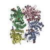
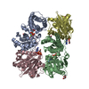
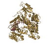
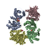
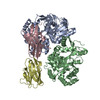
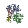
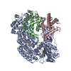
| Entry | Database: PDB / ID: 4g1t | ||||||
|---|---|---|---|---|---|---|---|
| Title | Crystal structure of interferon-stimulated gene 54 | ||||||
 Components Components | Interferon-induced protein with tetratricopeptide repeats 2 | ||||||
 Keywords Keywords | ANTIVIRAL PROTEIN / ISG / all alpha helix / antivirus | ||||||
| Function / homology |  Function and homology information Function and homology informationapoptotic mitochondrial changes / negative regulation of protein binding / antiviral innate immune response / response to virus / Interferon alpha/beta signaling / defense response to virus / positive regulation of apoptotic process / endoplasmic reticulum / RNA binding / cytoplasm / cytosol Similarity search - Function | ||||||
| Biological species |  Homo sapiens (human) Homo sapiens (human) | ||||||
| Method |  X-RAY DIFFRACTION / SYNCHROTRON / SAD / Resolution: 2.8 Å X-RAY DIFFRACTION / SYNCHROTRON / SAD / Resolution: 2.8 Å | ||||||
 Authors Authors | Yang, Z. / Liang, H. / Zhou, Q. / Li, Y. / Chen, H. / Ye, W. / Chen, D. / Fleming, J. / Shu, H. / Liu, Y. | ||||||
 Citation Citation | Journal: Cell Res. / Year: 2012 Title: Crystal structure of ISG54 reveals a novel RNA binding structure and potential functional mechanisms. Authors: Yang, Z. / Liang, H. / Zhou, Q. / Li, Y. / Chen, H. / Ye, W. / Chen, D. / Fleming, J. / Shu, H. / Liu, Y. | ||||||
| History |
|
- Structure visualization
| Structure viewer | Molecule: MolmilJmol/JSmol |
|---|
- Downloads & links
-Download
| PDBx/mmCIF format | 4g1t.cif.gz | 177.7 KB | Display | PDBx/mmCIF format |
|---|---|---|---|---|
| PDB format | pdb4g1t.ent.gz | 141.7 KB | Display | PDB format |
| PDBx/mmJSON format | 4g1t.json.gz | Tree view | PDBx/mmJSON format | |
| Others |  Other downloads Other downloads |
-Validation report
| Arichive directory | https://data.pdbj.org/pub/pdb/validation_reports/g1/4g1tftp://data.pdbj.org/pub/pdb/validation_reports/g1/4g1t | HTTPS FTP |
|---|
-Related structure data
| Similar structure data |
|---|


-Links
 PDBj
PDBj


- Assembly
| Deposited unit | 
| ||||||||
|---|---|---|---|---|---|---|---|---|---|
| 1 |
| ||||||||
| Unit cell |
|
-Components
| #1: Protein | Mass: 54730.242 Da / Num. of mol.: 2 Source method: isolated from a genetically manipulated source Source: (gene. exp.) Homo sapiens (human) / Gene: IFIT2, G10P2, IFI54 / Production host:  #2: Water | ChemComp-HOH / |  Mass: 18.015 Da / Num. of mol.: 23 / Source method: isolated from a natural source / Formula: H2O Mass: 18.015 Da / Num. of mol.: 23 / Source method: isolated from a natural source / Formula: H2OSequence details | NATURAL VALIANT D -> E AT THIS POSITION | |
|---|
-Experimental details
-Experiment
| Experiment | Method: X-RAY DIFFRACTION / Number of used crystals: 2 |
|---|
- Sample preparation
| Crystal | Density Matthews: 2.71 Å3/Da / Density % sol: 54.66 % |
|---|---|
| Crystal grow | Temperature: 289 K / Method: vapor diffusion, hanging drop / pH: 7.4 Details: PEG, pH 7.4, VAPOR DIFFUSION, HANGING DROP, temperature 289K |
-Data collection
| Diffraction |
| |||||||||||||||||||||||||||||||||||||||||||||||||||||||||||||||
|---|---|---|---|---|---|---|---|---|---|---|---|---|---|---|---|---|---|---|---|---|---|---|---|---|---|---|---|---|---|---|---|---|---|---|---|---|---|---|---|---|---|---|---|---|---|---|---|---|---|---|---|---|---|---|---|---|---|---|---|---|---|---|---|---|
| Diffraction source |
| |||||||||||||||||||||||||||||||||||||||||||||||||||||||||||||||
| Detector |
| |||||||||||||||||||||||||||||||||||||||||||||||||||||||||||||||
| Radiation |
| |||||||||||||||||||||||||||||||||||||||||||||||||||||||||||||||
| Radiation wavelength |
| |||||||||||||||||||||||||||||||||||||||||||||||||||||||||||||||
| Reflection | Resolution: 2.8→40 Å / Num. all: 29758 / Num. obs: 29706 / % possible obs: 99 % / Observed criterion σ(F): 4.4 / Observed criterion σ(I): 121.7 / Biso Wilson estimate: 63.88 Å2 | |||||||||||||||||||||||||||||||||||||||||||||||||||||||||||||||
| Reflection shell |
|


- Processing
| Software |
| ||||||||||||||||||||||||||||||||||||||||||||||||||||||||||||||||||||||||||||||||||||
|---|---|---|---|---|---|---|---|---|---|---|---|---|---|---|---|---|---|---|---|---|---|---|---|---|---|---|---|---|---|---|---|---|---|---|---|---|---|---|---|---|---|---|---|---|---|---|---|---|---|---|---|---|---|---|---|---|---|---|---|---|---|---|---|---|---|---|---|---|---|---|---|---|---|---|---|---|---|---|---|---|---|---|---|---|---|
| Refinement | Method to determine structure: SAD / Resolution: 2.8→39.572 Å / Occupancy max: 1 / Occupancy min: 1 / FOM work R set: 0.7453 / SU ML: 0.41 / σ(F): 0 / Phase error: 31.12 / Stereochemistry target values: ML
| ||||||||||||||||||||||||||||||||||||||||||||||||||||||||||||||||||||||||||||||||||||
| Solvent computation | Shrinkage radii: 0.95 Å / VDW probe radii: 1.2 Å / Solvent model: FLAT BULK SOLVENT MODEL / Bsol: 34.97 Å2 / ksol: 0.301 e/Å3 | ||||||||||||||||||||||||||||||||||||||||||||||||||||||||||||||||||||||||||||||||||||
| Displacement parameters | Biso max: 172.71 Å2 / Biso mean: 70.293 Å2 / Biso min: 20 Å2
| ||||||||||||||||||||||||||||||||||||||||||||||||||||||||||||||||||||||||||||||||||||
| Refinement step | Cycle: LAST / Resolution: 2.8→39.572 Å
| ||||||||||||||||||||||||||||||||||||||||||||||||||||||||||||||||||||||||||||||||||||
| Refine LS restraints |
| ||||||||||||||||||||||||||||||||||||||||||||||||||||||||||||||||||||||||||||||||||||
| LS refinement shell | Refine-ID: X-RAY DIFFRACTION / Total num. of bins used: 11
|
