- PDB-4q5v: Crystal structure of the catalytic core of human DNA polymerase a... -
+
Open data
ID or keywords:
Loading...
-
Basic information
Entry
Database: PDB / ID: 4q5v
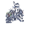
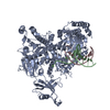
Title
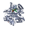
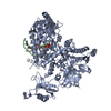
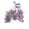
Crystal structure of the catalytic core of human DNA polymerase alpha in ternary complex with an RNA-primed DNA template and aphidicolin
Components
DNA polymerase alpha catalytic subunit
DNA template
RNA primer
Keywords
TRANSFERASE/DNA/RNA / B-family DNA polymerase / DNA replication / TRANSFERASE-DNA-RNA complex
Function / homology
Function and homology information
DNA replication initiation / Inhibition of replication initiation of damaged DNA by RB1/E2F1 / alpha DNA polymerase:primase complex / regulation of type I interferon production / Telomere C-strand synthesis initiation / Polymerase switching / Processive synthesis on the lagging strand / Removal of the Flap Intermediate / lagging strand elongation / DNA replication, synthesis of primer ...DNA replication initiation / Inhibition of replication initiation of damaged DNA by RB1/E2F1 / alpha DNA polymerase:primase complex / regulation of type I interferon production / Telomere C-strand synthesis initiation / Polymerase switching / Processive synthesis on the lagging strand / Removal of the Flap Intermediate / lagging strand elongation / DNA replication, synthesis of primer / mitotic DNA replication initiation / Polymerase switching on the C-strand of the telomere / DNA strand elongation involved in DNA replication / leading strand elongation / G1/S-Specific Transcription / DNA synthesis involved in DNA repair / DNA replication origin binding / Activation of the pre-replicative complex / DNA replication initiation / Defective pyroptosis / double-strand break repair via nonhomologous end joining / nuclear matrix / nuclear envelope / single-stranded DNA binding / DNA-directed DNA polymerase / DNA-directed DNA polymerase activity / DNA replication / nucleotide binding / DNA repair / chromatin binding / protein kinase binding / chromatin / nucleolus / DNA binding / zinc ion binding / nucleoplasm / nucleus / cytosol Similarity search - Function
OB fold (Dihydrolipoamide Acetyltransferase, E2P) - #730 / Alpha-Beta Plaits - #2820 / B family DNA polymerase, thumb domain / DNA polymerase alpha catalytic subunit, N-terminal domain / DNA polymerase alpha, zinc finger domain superfamily / DNA Polymerase alpha zinc finger / DNA polymerase alpha subunit p180 N terminal / Zinc finger, DNA-directed DNA polymerase, family B, alpha / DNA polymerase alpha catalytic subunit, catalytic domain / DNA polymerase family B, thumb domain ...OB fold (Dihydrolipoamide Acetyltransferase, E2P) - #730 / Alpha-Beta Plaits - #2820 / B family DNA polymerase, thumb domain / DNA polymerase alpha catalytic subunit, N-terminal domain / DNA polymerase alpha, zinc finger domain superfamily / DNA Polymerase alpha zinc finger / DNA polymerase alpha subunit p180 N terminal / Zinc finger, DNA-directed DNA polymerase, family B, alpha / DNA polymerase alpha catalytic subunit, catalytic domain / DNA polymerase family B, thumb domain / DNA-directed DNA polymerase, family B, multifunctional domain / DNA-directed DNA polymerase, family B, conserved site / DNA polymerase family B signature. / DNA polymerase family B / Topoisomerase I; Chain A, domain 4 / DNA polymerase family B, exonuclease domain / DNA-directed DNA polymerase, family B, exonuclease domain / DNA polymerase, palm domain superfamily / DNA polymerase type-B family / DNA-directed DNA polymerase, family B / Ribonuclease H-like superfamily/Ribonuclease H / OB fold (Dihydrolipoamide Acetyltransferase, E2P) / Nucleotidyltransferase; domain 5 / Ribonuclease H superfamily / Ribonuclease H-like superfamily / Alpha-Beta Plaits / DNA/RNA polymerase superfamily / Beta Barrel / 2-Layer Sandwich / Orthogonal Bundle / Mainly Beta / Mainly Alpha / Alpha Beta Similarity search - Domain/homology
Chem-2ZE / DNA / DNA (> 10) / RNA / RNA (> 10) / DNA polymerase alpha catalytic subunit Similarity search - Component
A: DNA polymerase alpha catalytic subunit B: RNA primer C: DNA template E: DNA polymerase alpha catalytic subunit F: RNA primer G: DNA template hetero molecules
Mass: 18.015 Da / Num. of mol.: 151 / Source method: isolated from a natural source / Formula: H2O
-
Experimental details
-
Experiment
Experiment
Method: X-RAY DIFFRACTION / Number of used crystals: 1
-
Sample preparation
Crystal
Density Matthews: 3.08 Å3/Da / Density % sol: 60.1 %
Crystal grow
Temperature: 295 K / Method: vapor diffusion, sitting drop / pH: 6.5 Details: 100 mM KCl, 12.5 mM MgCl2, 25 mM Na-Cacodylate, pH 6.5, 6 % 2-propanol, 2 mM TCEP, VAPOR DIFFUSION, SITTING DROP, temperature 295 K
In the structure databanks used in Yorodumi, some data are registered as the other names, "COVID-19 virus" and "2019-nCoV". Here are the details of the virus and the list of structure data.
Jan 31, 2019. EMDB accession codes are about to change! (news from PDBe EMDB page)
EMDB accession codes are about to change! (news from PDBe EMDB page)
The allocation of 4 digits for EMDB accession codes will soon come to an end. Whilst these codes will remain in use, new EMDB accession codes will include an additional digit and will expand incrementally as the available range of codes is exhausted. The current 4-digit format prefixed with “EMD-” (i.e. EMD-XXXX) will advance to a 5-digit format (i.e. EMD-XXXXX), and so on. It is currently estimated that the 4-digit codes will be depleted around Spring 2019, at which point the 5-digit format will come into force.
The EM Navigator/Yorodumi systems omit the EMD- prefix.
Related info.:Q: What is EMD? / ID/Accession-code notation in Yorodumi/EM Navigator
Yorodumi is a browser for structure data from EMDB, PDB, SASBDB, etc.
This page is also the successor to EM Navigator detail page, and also detail information page/front-end page for Omokage search.
The word "yorodu" (or yorozu) is an old Japanese word meaning "ten thousand". "mi" (miru) is to see.
Related info.:EMDB / PDB / SASBDB / Comparison of 3 databanks / Yorodumi Search / Aug 31, 2016. New EM Navigator & Yorodumi / Yorodumi Papers / Jmol/JSmol / Function and homology information / Changes in new EM Navigator and Yorodumi
 Movie
Movie Controller

 Yorodumi
 Open data
 Basic information
 Components
 Keywords
 Function and homology information
 Homo sapiens (human)
 X-RAY DIFFRACTION /
  Authors
 Citation
 Structure visualization
 Downloads & links
 Other downloads
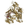


 PDBj
PDBj


 Assembly


 Spodoptera frugiperda (fall armyworm) / Strain (production host): SF-21 / References: UniProt: P09884, DNA-directed DNA polymerase
 Mass: 338.482 Da / Num. of mol.: 2 / Source method: obtained synthetically / Formula: C20H34O4
 Mass: 18.015 Da / Num. of mol.: 151 / Source method: isolated from a natural source / Formula: H2O
 Sample preparation
 / Beamline: 24-ID-C
 Processing