 Movie
Movie Controller

[English] 日本語
 Yorodumi
- PDB-4fkm: Structure of unliganded and reductively methylated FhuD2 from sta... -
+ Open data

- Basic information
| Entry | Database: PDB / ID: 4fkm | ||||||
|---|---|---|---|---|---|---|---|
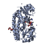
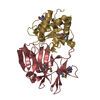
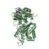
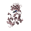
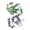
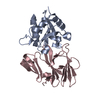
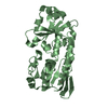
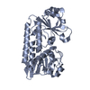
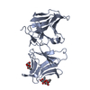
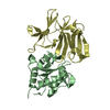
| Title | Structure of unliganded and reductively methylated FhuD2 from staphylococcus aureus | ||||||
 Components Components | Similar to ferric hydroxamate receptor 1 | ||||||
 Keywords Keywords | METAL BINDING PROTEIN / Class III Solute Binding Protein / membrane protein / transport of hydroxamate siderophores / Reductive methylation of lysyl residues | ||||||
| Function / homology |  Function and homology information Function and homology informationiron coordination entity transport / outer membrane-bounded periplasmic space / metal ion binding Similarity search - Function | ||||||
| Biological species |   Staphylococcus aureus (bacteria) Staphylococcus aureus (bacteria) | ||||||
| Method |  X-RAY DIFFRACTION / SYNCHROTRON / MAD / Resolution: 2.2 Å X-RAY DIFFRACTION / SYNCHROTRON / MAD / Resolution: 2.2 Å | ||||||
 Authors Authors | Podkowa, K.J. / Heinrichs, D.E. / Shilton, B.H. | ||||||
 Citation Citation | Journal: Biochemistry / Year: 2014 Title: Crystal and solution structure analysis of FhuD2 from Staphylococcus aureus in multiple unliganded conformations and bound to ferrioxamine-B. Authors: Podkowa, K.J. / Briere, L.A. / Heinrichs, D.E. / Shilton, B.H. | ||||||
| History |
|
- Structure visualization
| Structure viewer | Molecule: MolmilJmol/JSmol |
|---|
- Downloads & links
-Download
| PDBx/mmCIF format | 4fkm.cif.gz | 124.3 KB | Display | PDBx/mmCIF format |
|---|---|---|---|---|
| PDB format | pdb4fkm.ent.gz | 102.9 KB | Display | PDB format |
| PDBx/mmJSON format | 4fkm.json.gz | Tree view | PDBx/mmJSON format | |
| Others |  Other downloads Other downloads |
-Validation report
| Summary document | 4fkm_validation.pdf.gz | 475.2 KB | Display | wwPDB validaton report |
|---|---|---|---|---|
| Full document | 4fkm_full_validation.pdf.gz | 495.2 KB | Display | |
| Data in XML | 4fkm_validation.xml.gz | 28.9 KB | Display | |
| Data in CIF | 4fkm_validation.cif.gz | 37.3 KB | Display | |
| Arichive directory | https://data.pdbj.org/pub/pdb/validation_reports/fk/4fkmftp://data.pdbj.org/pub/pdb/validation_reports/fk/4fkm | HTTPS FTP |
-Related structure data


-Links
 PDBj
PDBj


- Assembly
| Deposited unit | 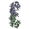
| ||||||||
|---|---|---|---|---|---|---|---|---|---|
| 1 | 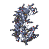
| ||||||||
| 2 | 
| ||||||||
| Unit cell |
|
-Components
| #1: Protein | Mass: 30878.389 Da / Num. of mol.: 2 / Fragment: UNP residues 44-301 Source method: isolated from a genetically manipulated source Details: expressed as GST fusion; GST removed using TEV protease Source: (gene. exp.) Staphylococcus aureus (bacteria) / Gene: SAV2284 / Plasmid: pGEX-FhuD2(delta)43 / Production host: #2: Water | ChemComp-HOH / |  Mass: 18.015 Da / Num. of mol.: 217 / Source method: isolated from a natural source / Formula: H2O Mass: 18.015 Da / Num. of mol.: 217 / Source method: isolated from a natural source / Formula: H2OHas protein modification | Y | |
|---|
-Experimental details
-Experiment
| Experiment | Method: X-RAY DIFFRACTION |
|---|
- Sample preparation
| Crystal | Density Matthews: 2.03 Å3/Da / Density % sol: 39.37 % |
|---|---|
| Crystal grow | Temperature: 291 K / Method: vapor diffusion, hanging drop / pH: 8.5 Details: 10 mg/mL protein concentration; 20% (w/v) PEG 3350, 25% (w/v) PEG 400, 100 mM MgCl2,100 mM Tris-HCl, pH 8.5, VAPOR DIFFUSION, HANGING DROP, temperature 291K |
-Data collection
| Diffraction | Mean temperature: 100 K | |||||||||||||||
|---|---|---|---|---|---|---|---|---|---|---|---|---|---|---|---|---|
| Diffraction source | Source: SYNCHROTRON / Site: NSLS  / Beamline: X8C / Wavelength: 1.0000, 0.9797, 0.9795, 0.9793 / Beamline: X8C / Wavelength: 1.0000, 0.9797, 0.9795, 0.9793 | |||||||||||||||
| Detector | Type: ADSC QUANTUM 4r / Detector: CCD / Date: Mar 21, 2005 | |||||||||||||||
| Radiation | Monochromator: Si(111) / Protocol: MAD / Monochromatic (M) / Laue (L): M / Scattering type: x-ray | |||||||||||||||
| Radiation wavelength |
| |||||||||||||||
| Reflection | Resolution: 2.2→60.3 Å / Num. all: 26093 / Num. obs: 26198 / % possible obs: 99.6 % / Observed criterion σ(F): 1 / Observed criterion σ(I): 1 / Redundancy: 3.8 % / Biso Wilson estimate: 27.7 Å2 / Rmerge(I) obs: 0.057 / Net I/σ(I): 16.1 | |||||||||||||||
| Reflection shell | Resolution: 2.2→2.32 Å / Redundancy: 3.6 % / Rmerge(I) obs: 0.255 / Mean I/σ(I) obs: 4.7 / Num. unique all: 3669 / % possible all: 98.3 |
- Processing
| Software |
| |||||||||||||||||||||||||
|---|---|---|---|---|---|---|---|---|---|---|---|---|---|---|---|---|---|---|---|---|---|---|---|---|---|---|
| Refinement | Method to determine structure: MAD / Resolution: 2.2→60 Å / Isotropic thermal model: Isotropic / Cross valid method: THROUGHOUT / σ(F): 0 / Stereochemistry target values: Engh & Huber
| |||||||||||||||||||||||||
| Displacement parameters | Biso mean: 29 Å2 | |||||||||||||||||||||||||
| Refinement step | Cycle: LAST / Resolution: 2.2→60 Å
| |||||||||||||||||||||||||
| Refine LS restraints |
|
