 Movie
Movie Controller


+ Open data

- Basic information
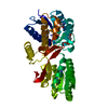
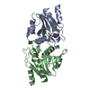
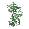
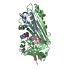
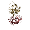
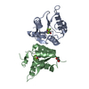
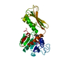
| Entry | Database: PDB / ID: 4fna | ||||||
|---|---|---|---|---|---|---|---|
| Title | Structure of unliganded FhuD2 from Staphylococcus Aureus | ||||||
 Components Components | Ferric hydroxamate receptor 2 | ||||||
 Keywords Keywords | METAL BINDING PROTEIN / Class III Solute Binding Protein / transport of hydroxamate siderophores / FhuCBG / membrane-bound | ||||||
| Function / homology |  Function and homology information Function and homology informationiron coordination entity transport / outer membrane-bounded periplasmic space Similarity search - Function | ||||||
| Biological species |   Staphylococcus aureus (bacteria) Staphylococcus aureus (bacteria) | ||||||
| Method |  X-RAY DIFFRACTION / SYNCHROTRON / MOLECULAR REPLACEMENT / Resolution: 3.5 Å X-RAY DIFFRACTION / SYNCHROTRON / MOLECULAR REPLACEMENT / Resolution: 3.5 Å | ||||||
 Authors Authors | Shilton, B.H. / Heinrichs, D.E. | ||||||
 Citation Citation | Journal: Biochemistry / Year: 2014 Title: Crystal and solution structure analysis of FhuD2 from Staphylococcus aureus in multiple unliganded conformations and bound to ferrioxamine-B. Authors: Podkowa, K.J. / Briere, L.A. / Heinrichs, D.E. / Shilton, B.H. | ||||||
| History |
|
- Structure visualization
| Structure viewer | Molecule: MolmilJmol/JSmol |
|---|
- Downloads & links
-Download
| PDBx/mmCIF format | 4fna.cif.gz | 160.1 KB | Display | PDBx/mmCIF format |
|---|---|---|---|---|
| PDB format | pdb4fna.ent.gz | 130.6 KB | Display | PDB format |
| PDBx/mmJSON format | 4fna.json.gz | Tree view | PDBx/mmJSON format | |
| Others |  Other downloads Other downloads |
-Validation report
| Arichive directory | https://data.pdbj.org/pub/pdb/validation_reports/fn/4fnaftp://data.pdbj.org/pub/pdb/validation_reports/fn/4fna | HTTPS FTP |
|---|
-Related structure data
| Related structure data | 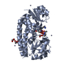 4filC 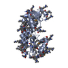 4fkmSC C: citing same article ( S: Starting model for refinement |
|---|---|
| Similar structure data |


-Links
 PDBj
PDBj


- Assembly
| Deposited unit | 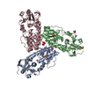
| ||||||||
|---|---|---|---|---|---|---|---|---|---|
| 1 | 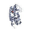
| ||||||||
| 2 | 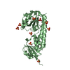
| ||||||||
| 3 | 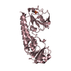
| ||||||||
| Unit cell |
|
-Components
| #1: Protein | Mass: 31617.119 Da / Num. of mol.: 3 / Fragment: UNP residues 25-302 Source method: isolated from a genetically manipulated source Details: expressed as GST fusion; GST removed using TEV protease Source: (gene. exp.) Staphylococcus aureus (bacteria) / Gene: fhud2 / Plasmid: pMTS57 / Production host: #2: Chemical | ChemComp-SO4 /   Mass: 96.063 Da / Num. of mol.: 13 / Source method: obtained synthetically / Formula: SO4 Mass: 96.063 Da / Num. of mol.: 13 / Source method: obtained synthetically / Formula: SO4 |
|---|
-Experimental details
-Experiment
| Experiment | Method: X-RAY DIFFRACTION |
|---|
- Sample preparation
| Crystal | Density Matthews: 4.62 Å3/Da / Density % sol: 73.37 % |
|---|---|
| Crystal grow | Temperature: 278 K / Method: vapor diffusion, sitting drop / pH: 3.5 Details: 0.1 M glycine, 3.5 to 3.8 M ammonium sulfate, pH 3.5, VAPOR DIFFUSION, SITTING DROP, temperature 278K |
-Data collection
| Diffraction | Mean temperature: 100 K |
|---|---|
| Diffraction source | Source: SYNCHROTRON / Site: NSLS  / Beamline: X8C / Wavelength: 1.1 Å / Beamline: X8C / Wavelength: 1.1 Å |
| Detector | Type: ADSC QUANTUM 4r / Detector: CCD / Date: Aug 28, 2002 |
| Radiation | Monochromator: Si(111) double crystal monochromator / Protocol: SINGLE WAVELENGTH / Monochromatic (M) / Laue (L): M / Scattering type: x-ray |
| Radiation wavelength | Wavelength: 1.1 Å / Relative weight: 1 |
| Reflection | Resolution: 3.5→24.714 Å / Num. all: 22693 / Num. obs: 22690 / % possible obs: 100 % / Redundancy: 12.9 % / Biso Wilson estimate: 141 Å2 / Rmerge(I) obs: 0.047 / Net I/σ(I): 14.7 |
| Reflection shell | Resolution: 3.5→3.63 Å / Rmerge(I) obs: 0.222 / % possible all: 100 |
- Processing
| Software |
| |||||||||||||||||||||||||||||||||||||||||||||||||||||||||||||||
|---|---|---|---|---|---|---|---|---|---|---|---|---|---|---|---|---|---|---|---|---|---|---|---|---|---|---|---|---|---|---|---|---|---|---|---|---|---|---|---|---|---|---|---|---|---|---|---|---|---|---|---|---|---|---|---|---|---|---|---|---|---|---|---|---|
| Refinement | Method to determine structure: MOLECULAR REPLACEMENT Starting model: PDB Entry 4FKM Resolution: 3.5→24.714 Å / SU ML: 0.38 / σ(F): 1.33 / Phase error: 31.76 / Stereochemistry target values: ML
| |||||||||||||||||||||||||||||||||||||||||||||||||||||||||||||||
| Solvent computation | Shrinkage radii: 0.9 Å / VDW probe radii: 1.11 Å / Solvent model: FLAT BULK SOLVENT MODEL | |||||||||||||||||||||||||||||||||||||||||||||||||||||||||||||||
| Displacement parameters | Biso mean: 124.9 Å2
| |||||||||||||||||||||||||||||||||||||||||||||||||||||||||||||||
| Refinement step | Cycle: LAST / Resolution: 3.5→24.714 Å
| |||||||||||||||||||||||||||||||||||||||||||||||||||||||||||||||
| Refine LS restraints |
| |||||||||||||||||||||||||||||||||||||||||||||||||||||||||||||||
| LS refinement shell |
|
