 Movie
Movie Controller


+ Open data

- Basic information
| Entry | Database: PDB / ID: 4etr | ||||||
|---|---|---|---|---|---|---|---|
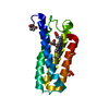
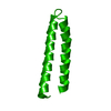
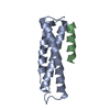
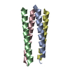
| Title | X-ray structure of PA2169 from Pseudomonas aeruginosa | ||||||
 Components Components | Putative uncharacterized protein | ||||||
 Keywords Keywords | UNKNOWN FUNCTION / DUF2383 / domain of unknown function / cytoplasmic | ||||||
| Function / homology |  Function and homology information Function and homology informationConserved hypothetical protein CHP02284 / Uncharacterised conserved protein UCP029477 / Domain of unknown function DUF2383 / Domain of unknown function (DUF2383) / Ferritin, core subunit, four-helix bundle / Ferritin / Ferritin-like / Ferritin-like superfamily / Up-down Bundle / Mainly Alpha Similarity search - Domain/homology | ||||||
| Biological species |   Pseudomonas aeruginosa (bacteria) Pseudomonas aeruginosa (bacteria) | ||||||
| Method |  X-RAY DIFFRACTION / SYNCHROTRON / MOLECULAR REPLACEMENT / Resolution: 2.25 Å X-RAY DIFFRACTION / SYNCHROTRON / MOLECULAR REPLACEMENT / Resolution: 2.25 Å | ||||||
 Authors Authors | Schnell, R. / Sandalova, T. / Lindqvist, Y. / Schneider, G. | ||||||
 Citation Citation | Journal: Acta Crystallogr.,Sect.F / Year: 2013 Title: The AEROPATH project targeting Pseudomonas aeruginosa: crystallographic studies for assessment of potential targets in early-stage drug discovery. Authors: Moynie, L. / Schnell, R. / McMahon, S.A. / Sandalova, T. / Boulkerou, W.A. / Schmidberger, J.W. / Alphey, M. / Cukier, C. / Duthie, F. / Kopec, J. / Liu, H. / Jacewicz, A. / Hunter, W.N. / ...Authors: Moynie, L. / Schnell, R. / McMahon, S.A. / Sandalova, T. / Boulkerou, W.A. / Schmidberger, J.W. / Alphey, M. / Cukier, C. / Duthie, F. / Kopec, J. / Liu, H. / Jacewicz, A. / Hunter, W.N. / Naismith, J.H. / Schneider, G. | ||||||
| History |
|
- Structure visualization
| Structure viewer | Molecule: MolmilJmol/JSmol |
|---|
- Downloads & links
-Download
| PDBx/mmCIF format | 4etr.cif.gz | 59.7 KB | Display | PDBx/mmCIF format |
|---|---|---|---|---|
| PDB format | pdb4etr.ent.gz | 43.6 KB | Display | PDB format |
| PDBx/mmJSON format | 4etr.json.gz | Tree view | PDBx/mmJSON format | |
| Others |  Other downloads Other downloads |
-Validation report
| Arichive directory | https://data.pdbj.org/pub/pdb/validation_reports/et/4etrftp://data.pdbj.org/pub/pdb/validation_reports/et/4etr | HTTPS FTP |
|---|
-Related structure data
| Related structure data | 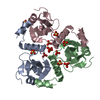 2xu8C 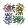 4avfC 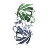 4avrC 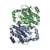 4avyC 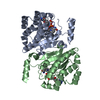 4b79C 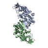 4b7cC 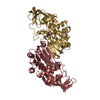 4b7xC 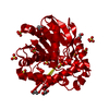 4b9aC 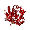 4b9eC 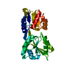 4es6C  4exaC 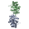 4exbC 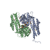 3fseS S: Starting model for refinement C: citing same article ( |
|---|---|
| Similar structure data |


-Links
 PDBj
PDBj


- Assembly
| Deposited unit | 
| ||||||||
|---|---|---|---|---|---|---|---|---|---|
| 1 | 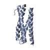
| ||||||||
| 2 | 
| ||||||||
| Unit cell |
|
-Components
| #1: Protein | Mass: 17152.094 Da / Num. of mol.: 2 / Fragment: PA2169 Source method: isolated from a genetically manipulated source Source: (gene. exp.) Pseudomonas aeruginosa (bacteria) / Strain: PAO1 / Gene: PA2169 / Plasmid: pET28a / Production host: #2: Water | ChemComp-HOH / |  Mass: 18.015 Da / Num. of mol.: 21 / Source method: isolated from a natural source / Formula: H2O Mass: 18.015 Da / Num. of mol.: 21 / Source method: isolated from a natural source / Formula: H2OHas protein modification | Y | |
|---|
-Experimental details
-Experiment
| Experiment | Method: X-RAY DIFFRACTION / Number of used crystals: 1 |
|---|
- Sample preparation
| Crystal | Density Matthews: 1.91 Å3/Da / Density % sol: 35.61 % |
|---|---|
| Crystal grow | Temperature: 293 K / Method: vapor diffusion, hanging drop / pH: 4.6 Details: 19% PEG10K, 100 mM Na-acetate pH 4.6, SrCl2 20mM, VAPOR DIFFUSION, HANGING DROP, temperature 293K |
-Data collection
| Diffraction | Mean temperature: 110 K |
|---|---|
| Diffraction source | Source: SYNCHROTRON / Site: ESRF  / Beamline: ID23-1 / Wavelength: 0.97625 Å / Beamline: ID23-1 / Wavelength: 0.97625 Å |
| Detector | Type: ADSC QUANTUM 315r / Detector: CCD / Date: Apr 17, 2009 / Details: mirrors |
| Radiation | Monochromator: Si 111 CHANNEL / Protocol: SINGLE WAVELENGTH / Monochromatic (M) / Laue (L): M / Scattering type: x-ray |
| Radiation wavelength | Wavelength: 0.97625 Å / Relative weight: 1 |
| Reflection | Resolution: 2.25→45.36 Å / Num. all: 12342 / Num. obs: 11947 / % possible obs: 96.8 % / Observed criterion σ(F): 2.6 / Observed criterion σ(I): 2.6 / Redundancy: 3.5 % / Biso Wilson estimate: 36.5 Å2 / Rmerge(I) obs: 0.092 / Rsym value: 0.092 / Net I/σ(I): 8.5 |
| Reflection shell | Resolution: 2.25→2.37 Å / Redundancy: 3.5 % / Rmerge(I) obs: 0.485 / Mean I/σ(I) obs: 2.6 / Num. unique all: 1726 / Rsym value: 0.485 / % possible all: 96.2 |
- Processing
| Software |
| |||||||||||||||||||||||||||||||||||||||||||||||||||||||||||||||||
|---|---|---|---|---|---|---|---|---|---|---|---|---|---|---|---|---|---|---|---|---|---|---|---|---|---|---|---|---|---|---|---|---|---|---|---|---|---|---|---|---|---|---|---|---|---|---|---|---|---|---|---|---|---|---|---|---|---|---|---|---|---|---|---|---|---|---|
| Refinement | Method to determine structure: MOLECULAR REPLACEMENT Starting model: PDB ENTRY 3FSE, trimmed to the 4 helix module, side chains removed Resolution: 2.25→45.35 Å / Cor.coef. Fo:Fc: 0.943 / Cor.coef. Fo:Fc free: 0.909 / SU B: 11.665 / SU ML: 0.265 / Isotropic thermal model: Isotropic / Cross valid method: THROUGHOUT / σ(I): 2.6 / ESU R: 0.375 / ESU R Free: 0.272 / Stereochemistry target values: MAXIMUM LIKELIHOOD / Details: HYDROGENS HAVE BEEN ADDED IN THE RIDING POSITIONS
| |||||||||||||||||||||||||||||||||||||||||||||||||||||||||||||||||
| Solvent computation | Ion probe radii: 0.8 Å / Shrinkage radii: 0.8 Å / VDW probe radii: 1.4 Å / Solvent model: BABINET MODEL WITH MASK | |||||||||||||||||||||||||||||||||||||||||||||||||||||||||||||||||
| Displacement parameters | Biso mean: 40.997 Å2
| |||||||||||||||||||||||||||||||||||||||||||||||||||||||||||||||||
| Refine analyze | Luzzati coordinate error free: 0.272 Å | |||||||||||||||||||||||||||||||||||||||||||||||||||||||||||||||||
| Refinement step | Cycle: LAST / Resolution: 2.25→45.35 Å
| |||||||||||||||||||||||||||||||||||||||||||||||||||||||||||||||||
| Refine LS restraints |
| |||||||||||||||||||||||||||||||||||||||||||||||||||||||||||||||||
| LS refinement shell | Resolution: 2.25→2.308 Å / Total num. of bins used: 20
|
