OXIDOREDUCTASE / oligomeric state / molecular switch / medium chain dehydrogenase / glucuronidation / MDR / Rossmann fold
Function / homology
Function and homology information
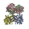
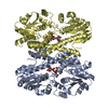
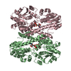
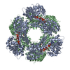
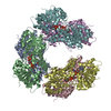
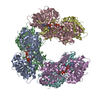
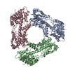
Formation of the active cofactor, UDP-glucuronate / UDP-glucose 6-dehydrogenase / UDP-glucose 6-dehydrogenase activity / UDP-glucuronate biosynthetic process / glycosaminoglycan biosynthetic process / chondroitin sulfate proteoglycan biosynthetic process / heparan sulfate proteoglycan biosynthetic process / gastrulation with mouth forming second / protein hexamerization / neuron development ...Formation of the active cofactor, UDP-glucuronate / UDP-glucose 6-dehydrogenase / UDP-glucose 6-dehydrogenase activity / UDP-glucuronate biosynthetic process / glycosaminoglycan biosynthetic process / chondroitin sulfate proteoglycan biosynthetic process / heparan sulfate proteoglycan biosynthetic process / gastrulation with mouth forming second / protein hexamerization / neuron development / NAD binding / extracellular exosome / nucleoplasm / identical protein binding / nucleus / cytosol Similarity search - Function
 Movie
Movie Controller


 Open data
 Basic information
 Components
 Keywords
 Function and homology information
 Homo sapiens (human)
 X-RAY DIFFRACTION /
  Authors
 Citation
 Structure visualization
 Downloads & links
 Other downloads
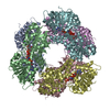
 PDBj
PDBj


 Assembly