 Movie
Movie Controller

[English] 日本語
 Yorodumi
- PDB-4dvr: Crystal structure of YU2 gp120 core in complex with Fab 48d and N... -
+ Open data

- Basic information
| Entry | Database: PDB / ID: 4dvr | ||||||
|---|---|---|---|---|---|---|---|
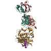
| Title | Crystal structure of YU2 gp120 core in complex with Fab 48d and NBD-557 | ||||||
 Components Components |
| ||||||
 Keywords Keywords | IMMUNE SYSTEM/TRANSCRIPTION INHIBITOR / HIV-1 gp120 / CD4 binding site / NBD-557 / IMMUNE SYSTEM-TRANSCRIPTION INHIBITOR complex | ||||||
| Function / homology |  Function and homology information Function and homology informationDectin-2 family / symbiont-mediated perturbation of host defense response / positive regulation of plasma membrane raft polarization / positive regulation of receptor clustering / host cell endosome membrane / clathrin-dependent endocytosis of virus by host cell / viral protein processing / fusion of virus membrane with host plasma membrane / fusion of virus membrane with host endosome membrane / viral envelope ...Dectin-2 family / symbiont-mediated perturbation of host defense response / positive regulation of plasma membrane raft polarization / positive regulation of receptor clustering / host cell endosome membrane / clathrin-dependent endocytosis of virus by host cell / viral protein processing / fusion of virus membrane with host plasma membrane / fusion of virus membrane with host endosome membrane / viral envelope / virion attachment to host cell / host cell plasma membrane / virion membrane / structural molecule activity / membrane Similarity search - Function | ||||||
| Biological species |  Human immunodeficiency virus type 1 group M subtype B Human immunodeficiency virus type 1 group M subtype B Homo sapiens (human) Homo sapiens (human) | ||||||
| Method |  X-RAY DIFFRACTION / SYNCHROTRON / MOLECULAR REPLACEMENT / Resolution: 2.5 Å X-RAY DIFFRACTION / SYNCHROTRON / MOLECULAR REPLACEMENT / Resolution: 2.5 Å | ||||||
 Authors Authors | Kwon, Y.D. / Kwong, P.D. | ||||||
 Citation Citation | Journal: Plos One / Year: 2014 Title: Crystal Structures of HIV-1 gp120 Envelope Glycoprotein in Complex with NBD Analogues That Target the CD4-Binding Site. Authors: Kwon, Y.D. / Lalonde, J.M. / Yang, Y. / Elban, M.A. / Sugawara, A. / Courter, J.R. / Jones, D.M. / Smith, A.B. / Debnath, A.K. / Kwong, P.D. | ||||||
| History |
|
- Structure visualization
| Structure viewer | Molecule: MolmilJmol/JSmol |
|---|
- Downloads & links
-Download
| PDBx/mmCIF format | 4dvr.cif.gz | 304.1 KB | Display | PDBx/mmCIF format |
|---|---|---|---|---|
| PDB format | pdb4dvr.ent.gz | 247.2 KB | Display | PDB format |
| PDBx/mmJSON format | 4dvr.json.gz | Tree view | PDBx/mmJSON format | |
| Others |  Other downloads Other downloads |
-Validation report
| Arichive directory | https://data.pdbj.org/pub/pdb/validation_reports/dv/4dvrftp://data.pdbj.org/pub/pdb/validation_reports/dv/4dvr | HTTPS FTP |
|---|
-Related structure data
| Related structure data |  4dvsC  4dvtC  4dvvC  4dvwC  4dvxC  1rzkS S: Starting model for refinement C: citing same article ( |
|---|---|
| Similar structure data |


-Links
 PDBj
PDBj


- Assembly
| Deposited unit | 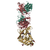
| ||||||||
|---|---|---|---|---|---|---|---|---|---|
| 1 |
| ||||||||
| Unit cell |
| ||||||||
| Details | monomer |
-Components
-Antibody , 2 types, 2 molecules LH
| #2: Antibody | Mass: 23092.562 Da / Num. of mol.: 1 Source method: isolated from a genetically manipulated source Source: (gene. exp.) Homo sapiens (human) / Production host: Homo sapiens (human) |
|---|---|
| #3: Antibody | Mass: 23386.137 Da / Num. of mol.: 1 Source method: isolated from a genetically manipulated source Source: (gene. exp.) Homo sapiens (human) / Production host: Homo sapiens (human) |
-Protein / Sugars , 2 types, 8 molecules G
| #1: Protein | Mass: 34918.633 Da / Num. of mol.: 1 / Fragment: unp residues 82-122; 199-297; 319-479 Source method: isolated from a genetically manipulated source Source: (gene. exp.) Human immunodeficiency virus type 1 group M subtype B (isolate YU-2)Strain: isolate YU-2 / Gene: env / Plasmid: pVRC8400 / Cell line (production host): HEK293F / Production host: Homo sapiens (human) / References: UniProt: P35961 |
|---|---|
| #4: Sugar | ChemComp-NAG /  Type: D-saccharide, beta linking / Mass: 221.208 Da / Num. of mol.: 7 Type: D-saccharide, beta linking / Mass: 221.208 Da / Num. of mol.: 7Source method: isolated from a genetically manipulated source Formula: C8H15NO6 |
-Non-polymers , 2 types, 114 molecules 

| #5: Chemical | ChemComp-0LY /  Mass: 382.295 Da / Num. of mol.: 1 / Source method: obtained synthetically / Formula: C17H24BrN3O2 Mass: 382.295 Da / Num. of mol.: 1 / Source method: obtained synthetically / Formula: C17H24BrN3O2 |
|---|---|
| #6: Water | ChemComp-HOH / Mass: 18.015 Da / Num. of mol.: 113 / Source method: isolated from a natural source / Formula: H2O |
-Details
| Has protein modification | Y |
|---|---|
| Sequence details | THE CRYSTALLIZED SEQUENCE WAS CONSTRUCTED BY DELETION OF N-TERMINAL 1-43, BY REPLACING THE V1/V2 ...THE CRYSTALLIZ |
-Experimental details
-Experiment
| Experiment | Method: X-RAY DIFFRACTION / Number of used crystals: 1 |
|---|
- Sample preparation
| Crystal | Density Matthews: 2.35 Å3/Da / Density % sol: 47.75 % |
|---|---|
| Crystal grow | Temperature: 293 K / Method: vapor diffusion, hanging drop / pH: 8.5 Details: 20% PEG3350, 5% iso-propanol, 0.1M Tris, pH 8.5, VAPOR DIFFUSION, HANGING DROP, temperature 293K |
-Data collection
| Diffraction | Mean temperature: 100 K |
|---|---|
| Diffraction source | Source: SYNCHROTRON / Site: APS  / Beamline: 22-ID / Wavelength: 1 Å / Beamline: 22-ID / Wavelength: 1 Å |
| Detector | Type: MAR scanner 300 mm plate / Detector: IMAGE PLATE / Date: Jun 12, 2008 |
| Radiation | Protocol: SINGLE WAVELENGTH / Monochromatic (M) / Laue (L): M / Scattering type: x-ray |
| Radiation wavelength | Wavelength: 1 Å / Relative weight: 1 |
| Reflection | Resolution: 2.5→49.4 Å / Num. all: 27379 / Num. obs: 24559 / % possible obs: 89.7 % / Observed criterion σ(F): 0 / Observed criterion σ(I): 0 / Redundancy: 4.6 % / Biso Wilson estimate: 56.8 Å2 / Rmerge(I) obs: 0.134 / Rsym value: 0.155 / Net I/σ(I): 11.04 |
| Reflection shell | Resolution: 2.5→2.54 Å / Redundancy: 1.8 % / Rmerge(I) obs: 0.497 / Mean I/σ(I) obs: 2.2 / Rsym value: 0.46 / % possible all: 62.9 |
- Processing
| Software |
| |||||||||||||||||||||||||||||||||||||||||||||||||||||||||||||||||||||||||||||||||||||||||||||||||||||||||||||||||||||||||||||||||||||||||||||||||||||||||||||||||||||||||||||||
|---|---|---|---|---|---|---|---|---|---|---|---|---|---|---|---|---|---|---|---|---|---|---|---|---|---|---|---|---|---|---|---|---|---|---|---|---|---|---|---|---|---|---|---|---|---|---|---|---|---|---|---|---|---|---|---|---|---|---|---|---|---|---|---|---|---|---|---|---|---|---|---|---|---|---|---|---|---|---|---|---|---|---|---|---|---|---|---|---|---|---|---|---|---|---|---|---|---|---|---|---|---|---|---|---|---|---|---|---|---|---|---|---|---|---|---|---|---|---|---|---|---|---|---|---|---|---|---|---|---|---|---|---|---|---|---|---|---|---|---|---|---|---|---|---|---|---|---|---|---|---|---|---|---|---|---|---|---|---|---|---|---|---|---|---|---|---|---|---|---|---|---|---|---|---|---|---|
| Refinement | Method to determine structure: MOLECULAR REPLACEMENT Starting model: pdb entry 1RZK Resolution: 2.5→49.4 Å / Occupancy max: 1 / Occupancy min: 0 / SU ML: 0.46 / σ(F): 1.42 / Phase error: 35.42 / Stereochemistry target values: ML
| |||||||||||||||||||||||||||||||||||||||||||||||||||||||||||||||||||||||||||||||||||||||||||||||||||||||||||||||||||||||||||||||||||||||||||||||||||||||||||||||||||||||||||||||
| Solvent computation | Shrinkage radii: 0.9 Å / VDW probe radii: 1.11 Å / Solvent model: FLAT BULK SOLVENT MODEL | |||||||||||||||||||||||||||||||||||||||||||||||||||||||||||||||||||||||||||||||||||||||||||||||||||||||||||||||||||||||||||||||||||||||||||||||||||||||||||||||||||||||||||||||
| Displacement parameters | Biso mean: 71.8165 Å2
| |||||||||||||||||||||||||||||||||||||||||||||||||||||||||||||||||||||||||||||||||||||||||||||||||||||||||||||||||||||||||||||||||||||||||||||||||||||||||||||||||||||||||||||||
| Refinement step | Cycle: LAST / Resolution: 2.5→49.4 Å
| |||||||||||||||||||||||||||||||||||||||||||||||||||||||||||||||||||||||||||||||||||||||||||||||||||||||||||||||||||||||||||||||||||||||||||||||||||||||||||||||||||||||||||||||
| Refine LS restraints |
| |||||||||||||||||||||||||||||||||||||||||||||||||||||||||||||||||||||||||||||||||||||||||||||||||||||||||||||||||||||||||||||||||||||||||||||||||||||||||||||||||||||||||||||||
| LS refinement shell |
| |||||||||||||||||||||||||||||||||||||||||||||||||||||||||||||||||||||||||||||||||||||||||||||||||||||||||||||||||||||||||||||||||||||||||||||||||||||||||||||||||||||||||||||||
| Refinement TLS params. | Method: refined / Refine-ID: X-RAY DIFFRACTION
| |||||||||||||||||||||||||||||||||||||||||||||||||||||||||||||||||||||||||||||||||||||||||||||||||||||||||||||||||||||||||||||||||||||||||||||||||||||||||||||||||||||||||||||||
| Refinement TLS group |
|
