Movie
Movie Controller

[English] 日本語
 Yorodumi
- PDB-4ck5: Pseudo-atomic model of microtubule-bound human kinesin-5 motor do... -
+ Open data

- Basic information
| Entry | Database: PDB / ID: 4ck5 | ||||||
|---|---|---|---|---|---|---|---|
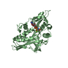
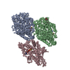
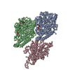
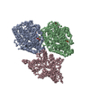
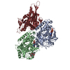
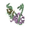
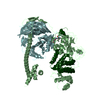
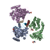
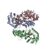
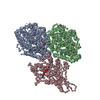
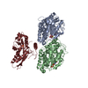
| Title | Pseudo-atomic model of microtubule-bound human kinesin-5 motor domain in the ADP state, based on cryo-electron microscopy experiment. | ||||||
 Components Components |
| ||||||
 Keywords Keywords | MOTOR PROTEIN / KINESINS / MICROTUBULES / MITOSIS / MECHANOCHEMISTRY | ||||||
| Function / homology |  Function and homology information Function and homology informationspindle elongation / regulation of mitotic centrosome separation / plus-end-directed microtubule motor activity / mitotic centrosome separation / Kinesins / positive regulation of axon guidance / spindle organization / microtubule motor activity / kinesin complex / COPI-dependent Golgi-to-ER retrograde traffic ...spindle elongation / regulation of mitotic centrosome separation / plus-end-directed microtubule motor activity / mitotic centrosome separation / Kinesins / positive regulation of axon guidance / spindle organization / microtubule motor activity / kinesin complex / COPI-dependent Golgi-to-ER retrograde traffic / microtubule-based movement / mitotic spindle assembly / microtubule-based process / MHC class II antigen presentation / mitotic spindle organization / sperm end piece / structural constituent of cytoskeleton / microtubule cytoskeleton organization / spindle / neuron migration / spindle pole / mitotic spindle / mitotic cell cycle / sperm principal piece / microtubule cytoskeleton / sperm midpiece / microtubule binding / microtubule / Hydrolases; Acting on acid anhydrides; Acting on GTP to facilitate cellular and subcellular movement / ciliary basal body / protein heterodimerization activity / cell division / hydrolase activity / GTPase activity / protein kinase binding / GTP binding / protein-containing complex / ATP binding / membrane / metal ion binding / nucleus / cytoplasm / cytosol Similarity search - Function | ||||||
| Biological species |  HOMO SAPIENS (human) HOMO SAPIENS (human) | ||||||
| Method | ELECTRON MICROSCOPY / single particle reconstruction / cryo EM / Resolution: 10 Å | ||||||
| Model type details | CA ATOMS ONLY, CHAIN A, B, C | ||||||
 Authors Authors | Goulet, A. / Major, J. / Jun, Y. / Gross, S. / Rosenfeld, S. / Moores, C. | ||||||
 Citation Citation | Journal: Proc Natl Acad Sci U S A / Year: 2014 Title: Comprehensive structural model of the mechanochemical cycle of a mitotic motor highlights molecular adaptations in the kinesin family. Authors: Adeline Goulet / Jennifer Major / Yonggun Jun / Steven P Gross / Steven S Rosenfeld / Carolyn A Moores /  Abstract: Kinesins are responsible for a wide variety of microtubule-based, ATP-dependent functions. Their motor domain drives these activities, but the molecular adaptations that specify these diverse and ...Kinesins are responsible for a wide variety of microtubule-based, ATP-dependent functions. Their motor domain drives these activities, but the molecular adaptations that specify these diverse and essential cellular activities are poorly understood. It has been assumed that the first identified kinesin--the transport motor kinesin-1--is the mechanistic paradigm for the entire superfamily, but accumulating evidence suggests otherwise. To address the deficits in our understanding of the molecular basis of functional divergence within the kinesin superfamily, we studied kinesin-5s, which are essential mitotic motors whose inhibition blocks cell division. Using cryo-electron microscopy and determination of structure at subnanometer resolution, we have visualized conformations of microtubule-bound human kinesin-5 motor domain at successive steps in its ATPase cycle. After ATP hydrolysis, nucleotide-dependent conformational changes in the active site are allosterically propagated into rotations of the motor domain and uncurling of the drug-binding loop L5. In addition, the mechanical neck-linker element that is crucial for motor stepping undergoes discrete, ordered displacements. We also observed large reorientations of the motor N terminus that indicate its importance for kinesin-5 function through control of neck-linker conformation. A kinesin-5 mutant lacking this N terminus is enzymatically active, and ATP-dependent neck-linker movement and motility are defective, although not ablated. All these aspects of kinesin-5 mechanochemistry are distinct from kinesin-1. Our findings directly demonstrate the regulatory role of the kinesin-5 N terminus in collaboration with the motor's structured neck-linker and highlight the multiple adaptations within kinesin motor domains that tune their mechanochemistries according to distinct functional requirements. | ||||||
| History |
|
- Structure visualization
| Movie |
Movie viewer |
|---|---|
| Structure viewer | Molecule: MolmilJmol/JSmol |
- Downloads & links
-Download
| PDBx/mmCIF format | 4ck5.cif.gz | 65.7 KB | Display | PDBx/mmCIF format |
|---|---|---|---|---|
| PDB format | pdb4ck5.ent.gz | 35.8 KB | Display | PDB format |
| PDBx/mmJSON format | 4ck5.json.gz | Tree view | PDBx/mmJSON format | |
| Others |  Other downloads Other downloads |
-Validation report
| Arichive directory | https://data.pdbj.org/pub/pdb/validation_reports/ck/4ck5ftp://data.pdbj.org/pub/pdb/validation_reports/ck/4ck5 | HTTPS FTP |
|---|
-Related structure data
| Related structure data | 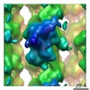 2537MC 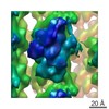 2533C 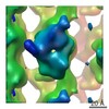 2534C  2535C  2536C  2538C  2539C  2540C  2541C  2542C 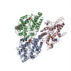 4ck6C  4ck7C C: citing same article ( M: map data used to model this data |
|---|---|
| Similar structure data |
-Links
 PDBj
PDBj


- Assembly
| Deposited unit | 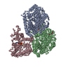
|
|---|---|
| 1 |
|
-Components
-Protein , 3 types, 3 molecules ABC
| #1: Protein | Mass: 50236.352 Da / Num. of mol.: 1 / Source method: isolated from a natural source / Source: (natural) |
|---|---|
| #2: Protein | Mass: 49907.770 Da / Num. of mol.: 1 / Source method: isolated from a natural source / Source: (natural) |
| #3: Protein | Mass: 41673.105 Da / Num. of mol.: 1 / Fragment: MOTOR DOMAIN, RESIDUES 1-367 / Mutation: YES Source method: isolated from a genetically manipulated source Source: (gene. exp.) HOMO SAPIENS (human) / Plasmid: PET21A / Production host:  |
-Non-polymers , 5 types, 5 molecules 


| #4: Chemical | ChemComp-GTP /  Mass: 523.180 Da / Num. of mol.: 1 / Source method: obtained synthetically / Formula: C10H16N5O14P3 / Comment: GTP, energy-carrying molecule*YM Mass: 523.180 Da / Num. of mol.: 1 / Source method: obtained synthetically / Formula: C10H16N5O14P3 / Comment: GTP, energy-carrying molecule*YM |
|---|---|
| #5: Chemical | ChemComp-GDP /  Type: RNA linking / Mass: 443.201 Da / Num. of mol.: 1 / Source method: obtained synthetically / Formula: C10H15N5O11P2 / Comment: GDP, energy-carrying molecule*YM Type: RNA linking / Mass: 443.201 Da / Num. of mol.: 1 / Source method: obtained synthetically / Formula: C10H15N5O11P2 / Comment: GDP, energy-carrying molecule*YM |
| #6: Chemical | ChemComp-TA1 /  Mass: 853.906 Da / Num. of mol.: 1 / Source method: obtained synthetically / Formula: C47H51NO14 / Comment: medication, chemotherapy*YM Mass: 853.906 Da / Num. of mol.: 1 / Source method: obtained synthetically / Formula: C47H51NO14 / Comment: medication, chemotherapy*YM |
| #7: Chemical | ChemComp-MG /  Mass: 24.305 Da / Num. of mol.: 1 / Source method: obtained synthetically / Formula: Mg Mass: 24.305 Da / Num. of mol.: 1 / Source method: obtained synthetically / Formula: Mg |
| #8: Chemical | ChemComp-ADP /  Mass: 427.201 Da / Num. of mol.: 1 / Source method: obtained synthetically / Formula: C10H15N5O10P2 / Comment: ADP, energy-carrying molecule*YM Mass: 427.201 Da / Num. of mol.: 1 / Source method: obtained synthetically / Formula: C10H15N5O10P2 / Comment: ADP, energy-carrying molecule*YM |
-Details
| Sequence details | CYS-LITE MUTANT CONTAINING |
|---|
-Experimental details
-Experiment
| Experiment | Method: ELECTRON MICROSCOPY |
|---|---|
| EM experiment | Aggregation state: PARTICLE / 3D reconstruction method: single particle reconstruction |
- Sample preparation
| Component | Name: microtubule-bound human kinesin-5 motor domain in the ADP state Type: COMPLEX Details: 13-PROTOFILAMENT MICROTUBULE- BOUND HUMAN KINESIN-5 MOTOR DOMAIN IN PRESENCE OF ADP |
|---|---|
| Buffer solution | Name: 20 MM PIPES, 5 MM MGCL2, 1 MM EGTA, 10 MM ADP / pH: 6.8 / Details: 20 MM PIPES, 5 MM MGCL2, 1 MM EGTA, 10 MM ADP |
| Specimen | Embedding applied: NO / Shadowing applied: NO / Staining applied: NO / Vitrification applied: YES |
| Vitrification | Instrument: FEI VITROBOT MARK I / Cryogen name: ETHANE |
- Electron microscopy imaging
| Experimental equipment |  Model: Tecnai F20 / Image courtesy: FEI Company |
|---|---|
| Microscopy | Model: FEI TECNAI F20 / Date: Oct 20, 2012 |
| Electron gun | Electron source:  FIELD EMISSION GUN / Accelerating voltage: 200 kV / Illumination mode: FLOOD BEAM FIELD EMISSION GUN / Accelerating voltage: 200 kV / Illumination mode: FLOOD BEAM |
| Electron lens | Mode: BRIGHT FIELD / Nominal magnification: 68000 X / Nominal defocus max: 2400 nm / Nominal defocus min: 700 nm / Cs: 2 mm |
| Specimen holder | Temperature: 90 K |
| Image recording | Electron dose: 18 e/Å2 / Film or detector model: GATAN ULTRASCAN 4000 (4k x 4k) |
| Image scans | Num. digital images: 160 |
- Processing
| EM software |
| |||||||||||||||||||||
|---|---|---|---|---|---|---|---|---|---|---|---|---|---|---|---|---|---|---|---|---|---|---|
| CTF correction | Details: FREALIGN | |||||||||||||||||||||
| Symmetry | Point symmetry: C1 (asymmetric) | |||||||||||||||||||||
| 3D reconstruction | Resolution: 10 Å / Num. of particles: 9615 / Actual pixel size: 2.2 Å Details: TO ACCOUNT FOR THE FACT THAT HELIX ALPHA4 IS LONGER IN OUR RECONSTRUCTION THAN IN THE AVAILABLE ADP CRYSTAL STRUCTURES, WE MODELLED 7 HELICAL TURNS OF HELIX ALPHA4 ( RESIDUES D279-E304), AS ...Details: TO ACCOUNT FOR THE FACT THAT HELIX ALPHA4 IS LONGER IN OUR RECONSTRUCTION THAN IN THE AVAILABLE ADP CRYSTAL STRUCTURES, WE MODELLED 7 HELICAL TURNS OF HELIX ALPHA4 ( RESIDUES D279-E304), AS OBSERVED IN THE ENTRY 3HQD. THE N- TERMINAL SEQUENCE WAS MODELLED USING MODELLER. SUBMISSION BASED ON EXPERIMENTAL DATA FROM EMDB EMD-2537. (DEPOSITION ID: 12202). Symmetry type: POINT | |||||||||||||||||||||
| Atomic model building | Protocol: FLEXIBLE FIT / Space: REAL / Target criteria: Cross-correlation coefficient / Details: METHOD--FLEXIBLE | |||||||||||||||||||||
| Atomic model building |
| |||||||||||||||||||||
| Refinement | Highest resolution: 10 Å | |||||||||||||||||||||
| Refinement step | Cycle: LAST / Highest resolution: 10 Å
|