 Movie
Movie Controller


+ Open data

- Basic information
| Entry | Database: PDB / ID: 1ia0 | ||||||
|---|---|---|---|---|---|---|---|
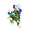
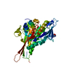
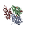
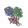
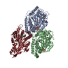
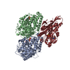
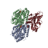
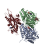
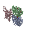
| Title | KIF1A HEAD-MICROTUBULE COMPLEX STRUCTURE IN ATP-FORM | ||||||
 Components Components |
| ||||||
 Keywords Keywords | TRANSPORT PROTEIN / tubulin / microtubule / KIF1A / FITTING OF X-RAY STRUCTURES INTO CRYO-EM RECONSTRUCTIONS | ||||||
| Function / homology |  Function and homology information Function and homology informationanterograde dendritic transport / interkinetic nuclear migration / anterograde synaptic vesicle transport / protein transport along microtubule / neuronal dense core vesicle membrane / dense core granule cytoskeletal transport / plus-end-directed kinesin ATPase / Kinesins / anterograde neuronal dense core vesicle transport / retrograde neuronal dense core vesicle transport ...anterograde dendritic transport / interkinetic nuclear migration / anterograde synaptic vesicle transport / protein transport along microtubule / neuronal dense core vesicle membrane / dense core granule cytoskeletal transport / plus-end-directed kinesin ATPase / Kinesins / anterograde neuronal dense core vesicle transport / retrograde neuronal dense core vesicle transport / regulation of dendritic spine development / COPI-dependent Golgi-to-ER retrograde traffic / MHC class II antigen presentation / regulation of dendritic spine morphogenesis / anterograde axonal transport / plus-end-directed microtubule motor activity / kinesin complex / neuronal dense core vesicle / vesicle-mediated transport / axon cytoplasm / dendrite cytoplasm / structural constituent of cytoskeleton / microtubule cytoskeleton organization / neuron migration / synaptic vesicle / mitotic cell cycle / presynapse / microtubule binding / microtubule / Hydrolases; Acting on acid anhydrides; Acting on GTP to facilitate cellular and subcellular movement / neuron projection / postsynapse / axon / hydrolase activity / neuronal cell body / GTPase activity / dendrite / GTP binding / perinuclear region of cytoplasm / glutamatergic synapse / ATP hydrolysis activity / protein-containing complex / ATP binding / metal ion binding / identical protein binding / cytoplasm Similarity search - Function | ||||||
| Biological species |  | ||||||
| Method | ELECTRON MICROSCOPY / helical reconstruction / cryo EM / Resolution: 15 Å | ||||||
 Authors Authors | Kikkawa, M. / Sablin, E.P. / Okada, Y. / Yajima, H. / Fletterick, R.J. / Hirokawa, N. | ||||||
 Citation Citation | Journal: Nature / Year: 2001 Title: Switch-based mechanism of kinesin motors. Authors: M Kikkawa / E P Sablin / Y Okada / H Yajima / R J Fletterick / N Hirokawa /  Abstract: Kinesin motors are specialized enzymes that use hydrolysis of ATP to generate force and movement along their cellular tracks, the microtubules. Although numerous biochemical and biophysical studies ...Kinesin motors are specialized enzymes that use hydrolysis of ATP to generate force and movement along their cellular tracks, the microtubules. Although numerous biochemical and biophysical studies have accumulated much data that link microtubule-assisted ATP hydrolysis to kinesin motion, the structural view of kinesin movement remains unclear. This study of the monomeric kinesin motor KIF1A combines X-ray crystallography and cryo-electron microscopy, and allows analysis of force-generating conformational changes at atomic resolution. The motor is revealed in its two functionally critical states-complexed with ADP and with a non-hydrolysable analogue of ATP. The conformational change observed between the ADP-bound and the ATP-like structures of the KIF1A catalytic core is modular, extends to all kinesins and is similar to the conformational change used by myosin motors and G proteins. Docking of the ADP-bound and ATP-like crystallographic models of KIF1A into the corresponding cryo-electron microscopy maps suggests a rationale for the plus-end directional bias associated with the kinesin catalytic core. #1: Journal: Cell(Cambridge,Mass.) / Year: 2000Title: 15 Angstrom Resolution Model of the Monomeric Kinesin Motor, KIF1A Authors: Kikkawa, M. / Okada, Y. / Hirokawa, N. | ||||||
| History |
| ||||||
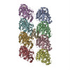
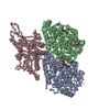
| Remark 300 | BIOMOLECULE: 1 THIS ENTRY CONTAINS THE CRYSTALLOGRAPHIC ASYMMETRIC UNIT WHICH CONSISTS OF 3 CHAIN(S) ...BIOMOLECULE: 1 THIS ENTRY CONTAINS THE CRYSTALLOGRAPHIC ASYMMETRIC UNIT WHICH CONSISTS OF 3 CHAIN(S). SEE REMARK 350 FOR INFORMATION ON GENERATING THE BIOLOGICAL MOLECULE(S). THE COMPLEX PARTICLE HAS A HELICAL SYMMETRY WITH 15 PROTOFILAMENTS. | ||||||
| Remark 350 | GENERATING THE BIOMOLECULE COORDINATES FOR A COMPLETE MULTIMER REPRESENTING THE KNOWN BIOLOGICALLY ...GENERATING THE BIOMOLECULE COORDINATES FOR A COMPLETE MULTIMER REPRESENTING THE KNOWN BIOLOGICALLY SIGNIFICANT OLIGOMERIZATION STATE OF THE MOLECULE CAN BE GENERATED BY APPLYING BIOMT TRANSFORMATIONS GIVEN BELOW. BOTH NON-CRYSTALLOGRAPHIC AND CRYSTALLOGRAPHIC OPERATIONS ARE GIVEN. LET P(0,0) = COORDINATES OF ANY ATOM AS LISTED IN ENTRY 1. BIOMT1 1 0.914728 -0.404068 0.000000 0.000000 BIOMT2 1 0.404068 0.914728 0.000000 0.000000 BIOMT3 1 0.000000 0.000000 1.000000 10.592334 APPLY FOLLOWING OPERATOR REPETITIVELY TO GENERATE SHALLOW HELIX. P(N+1,0) = BIOMT 1 * P(N,0) 2. BIOMT1 2 0.999042 0.043771 0.000000 0.000000 BIOMT2 2 -0.043771 0.999042 0.000000 0.000000 BIOMT3 2 0.000000 0.000000 1.000000 79.442509 THEN APPLY FOLLOWING OPERATOR REPETITIVELY TO GENERATE WHOLE KIF1A-MICROTUBULE COMPLEX. P(N,M+1) = BIOMT 2 * P(N,M) |
- Structure visualization
| Movie |
Movie viewer |
|---|---|
| Structure viewer | Molecule: MolmilJmol/JSmol |
- Downloads & links
-Download
| PDBx/mmCIF format | 1ia0.cif.gz | 258.4 KB | Display | PDBx/mmCIF format |
|---|---|---|---|---|
| PDB format | pdb1ia0.ent.gz | 177.7 KB | Display | PDB format |
| PDBx/mmJSON format | 1ia0.json.gz | Tree view | PDBx/mmJSON format | |
| Others |  Other downloads Other downloads |
-Validation report
| Arichive directory | https://data.pdbj.org/pub/pdb/validation_reports/ia/1ia0ftp://data.pdbj.org/pub/pdb/validation_reports/ia/1ia0 | HTTPS FTP |
|---|
-Related structure data


-Links
 PDBj
PDBj


- Assembly
| Deposited unit | 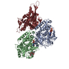
|
|---|---|
| 1 |
|
-Components
-Protein , 3 types, 3 molecules ABK
| #1: Protein | Mass: 50121.266 Da / Num. of mol.: 1 / Source method: isolated from a natural source / Source: (natural) |
|---|---|
| #2: Protein | Mass: 49907.770 Da / Num. of mol.: 1 / Source method: isolated from a natural source / Source: (natural) |
| #3: Protein | Mass: 44302.855 Da / Num. of mol.: 1 / Fragment: MOTOR DOMAIN / Mutation: P202A Source method: isolated from a genetically manipulated source Source: (gene. exp.)  |
-Non-polymers , 5 types, 5 molecules 


| #4: Chemical | ChemComp-GTP /  Mass: 523.180 Da / Num. of mol.: 1 / Source method: obtained synthetically / Formula: C10H16N5O14P3 / Comment: GTP, energy-carrying molecule*YM Mass: 523.180 Da / Num. of mol.: 1 / Source method: obtained synthetically / Formula: C10H16N5O14P3 / Comment: GTP, energy-carrying molecule*YM |
|---|---|
| #5: Chemical | ChemComp-GDP /  Type: RNA linking / Mass: 443.201 Da / Num. of mol.: 1 / Source method: obtained synthetically / Formula: C10H15N5O11P2 / Comment: GDP, energy-carrying molecule*YM Type: RNA linking / Mass: 443.201 Da / Num. of mol.: 1 / Source method: obtained synthetically / Formula: C10H15N5O11P2 / Comment: GDP, energy-carrying molecule*YM |
| #6: Chemical | ChemComp-TXL /  Mass: 807.879 Da / Num. of mol.: 1 / Source method: obtained synthetically / Formula: C43H53NO14 Mass: 807.879 Da / Num. of mol.: 1 / Source method: obtained synthetically / Formula: C43H53NO14 |
| #7: Chemical | ChemComp-MG /  Mass: 24.305 Da / Num. of mol.: 1 / Source method: obtained synthetically / Formula: Mg Mass: 24.305 Da / Num. of mol.: 1 / Source method: obtained synthetically / Formula: Mg |
| #8: Chemical | ChemComp-ACP /  Mass: 505.208 Da / Num. of mol.: 1 / Source method: obtained synthetically / Formula: C11H18N5O12P3 / Comment: AMP-PCP, energy-carrying molecule analogue*YM Mass: 505.208 Da / Num. of mol.: 1 / Source method: obtained synthetically / Formula: C11H18N5O12P3 / Comment: AMP-PCP, energy-carrying molecule analogue*YM |
-Details
| Has protein modification | N |
|---|
-Experimental details
-Experiment
| Experiment | Method: ELECTRON MICROSCOPY |
|---|---|
| EM experiment | Aggregation state: FILAMENT / 3D reconstruction method: helical reconstruction |
- Sample preparation
| Component | Name: KIF1A HEAD DECORATED MICROTUBULE / Type: COMPLEX Details: TUBULIN WAS POLYMERIZED IN PEM BUFFER (PIPES 100 MM, PH 6.8, EGTA 1 MM, MGCL2, GTP 1MM, PACLITAXEL 10 MICRO M, 5% DMSO ) FOR 2 HRS AT 37C. A DROP OF THE POLYMERIZED MICROTUBULE WAS PLACED ON ...Details: TUBULIN WAS POLYMERIZED IN PEM BUFFER (PIPES 100 MM, PH 6.8, EGTA 1 MM, MGCL2, GTP 1MM, PACLITAXEL 10 MICRO M, 5% DMSO ) FOR 2 HRS AT 37C. A DROP OF THE POLYMERIZED MICROTUBULE WAS PLACED ON THE HOLEY CARBON FILM ON THE EM-GRID AND LEFT FOR 10 S IN THE HUMID CHAMBER. THE MICROTUBULE SOLUTION WAS THEN ABSORBED BY FILTER PAPER, AND A DROP OF THE C351 SOLUTION (0.2 MG/ML) IN THE ASSAY BUFFER (IMIDAZOLE 50 MM, MG-ACETATE 5 MM, EGTA 1 MM, K-ACETATE 50 MM, DTT 10 MM, PACLITAXEL 10 MICRO M) WAS PUT ON THE GRID. IMMEDIATELY AFTER THE ABSORPTION OF THE DROP, THE GRID WAS PLUNGED INTO LIQUID ETHANE (-185C). THE SAMPLE CONSISTS OF A KIF1A-DECORATED MICROTUBULE. THE EXPERIMENT DESCRIBES A MICROTUBULE-RELATED PROCESS. |
|---|---|
| Buffer solution | Name: buf1 / pH: 7.4 Details: IMIDAZOLE 50 MM, MG-ACETATE 5 MM, EGTA 1 MM, K-ACETATE 50 MM, DTT 10 MM, PACLITAXEL 10 MICRO M |
| Specimen | Conc.: 0.2 mg/ml / Embedding applied: NO / Shadowing applied: NO / Staining applied: NO / Vitrification applied: YES |
| Specimen support | Details: HOLEY CARBON |
| Vitrification | Details: PLUNGED INTO ETHANE |
| Crystal grow | Temperature: 296 K / Method: polymerization of the microtubule / pH: 6.8 Details: pH 6.8, polymerization of the microtubule, temperature 296K |
- Electron microscopy imaging
| Microscopy | Model: JEOL 2010F / Date: Jul 1, 1998 |
|---|---|
| Electron gun | Electron source:  FIELD EMISSION GUN / Accelerating voltage: 200 kV / Illumination mode: FLOOD BEAM FIELD EMISSION GUN / Accelerating voltage: 200 kV / Illumination mode: FLOOD BEAM |
| Electron lens | Mode: BRIGHT FIELD / Nominal magnification: 40000 X / Nominal defocus max: 3000 nm / Nominal defocus min: 1700 nm / Cs: 3.3 mm |
| Specimen holder | Temperature: 110 K / Tilt angle max: 2 ° / Tilt angle min: 0 ° |
| Image recording | Electron dose: 10 e/Å2 / Film or detector model: KODAK SO-163 FILM |
| Image scans | Num. digital images: 130 |
- Processing
| 3D reconstruction | Resolution: 15 Å / Num. of particles: 37000 / Nominal pixel size: 2.5 Å Details: THREE-DIMENSIONAL RECONSTRUCTION METHOD: FOURIER-BESSEL-TRANSFORM (DEROSIER AND MOORE, 1970 J.MOL.BIOL. 52, 355-369; BEROUKHIM AND UNWIN, 1997 ULTRAMICROSCOPY 70, 57-81). Software used: MRC ...Details: THREE-DIMENSIONAL RECONSTRUCTION METHOD: FOURIER-BESSEL-TRANSFORM (DEROSIER AND MOORE, 1970 J.MOL.BIOL. 52, 355-369; BEROUKHIM AND UNWIN, 1997 ULTRAMICROSCOPY 70, 57-81). Software used: MRC PROGRAMS. EFFECTIVE RESOLUTION OF THE RECONSTRUCTION: THE RESOLUTION OF THE FINAL RECONSTRUCTED DENSITY WAS DETERMINED TO BE AT LEAST 15 ANGSTROMS, AS MEASURED BY RANDOMLY SPLITTING THE PARTICLES INTO TWO SETS AND CALCULATING THE FOURIER SHELL CORRELATIONS BETWEEN THE TWO INDEPENDENT SET. (HEEL, 1987, ULTRAMICROSCOPY 21, 95-100). 1IA0 is an atomic model of KIF1A head-microtubule complex, which contains KIF1A, alpha-tubulin, and beta-tubulin. Since alpha- and beta-tubulin were indistinguishable at 15A resolution, the assignment of alpha- and beta-tubulin was arbitrary when 1IA0 was deposited. Authors state that they were aware of the limitation and didn't mention about the assignment of alpha- and beta-tubulin in the relevant 2001 Nature paper. Therefore, the main conclusions in the paper are still hold true. However, subsequently the assignment of alpha- and beta-tubulin turned out to be wrong and should be swapped. For the correct assignment and better structures, please use 2HXF. This is based on higher resolution (10 ) structure, which was published in EMBO J. 2006. 25:4187-4194. Symmetry type: HELICAL | ||||||||||||
|---|---|---|---|---|---|---|---|---|---|---|---|---|---|
| Atomic model building | Protocol: RIGID BODY FIT / Space: REAL / Target criteria: MAXIMUM CORRELATION / Details: REFINEMENT PROTOCOL--RIGID BODY REFINEMENT | ||||||||||||
| Atomic model building | PDB-ID: 1TUB Accession code: 1TUB / Source name: PDB / Type: experimental model | ||||||||||||
| Refinement | Highest resolution: 15 Å Details: 1IA0 is a atomic model of KIF1A head-microtubule complex, which contains KIF1A, alpha-tubulin, and beta-tubulin. Since alpha- and beta-tubulin were indistinguishable at 15A resolution, the ...Details: 1IA0 is a atomic model of KIF1A head-microtubule complex, which contains KIF1A, alpha-tubulin, and beta-tubulin. Since alpha- and beta-tubulin were indistinguishable at 15A resolution, the assignment of alpha- and beta-tubulin was arbitrary when 1IA0 was deposited. Authors state that they were aware of the limitation and didn't mention about the assignment of alpha- and beta-tubulin in the relevant 2001 Nature paper. Therefore, the main conclusions in the paper are still hold true. However, subsequently the assignment of alpha- and beta-tubulin turned out to be wrong and should be swapped. For the correct assignment and better structures, please use 2HXF. This is based on higher resolution (10 angstrom) structure, which was published in EMBO J. 2006. 25:4187-4194. | ||||||||||||
| Refinement step | Cycle: LAST / Highest resolution: 15 Å
|
