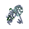
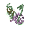
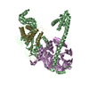
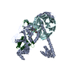
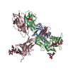
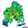
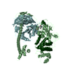
Mass: 72941.484 Da / Num. of mol.: 2 Source method: isolated from a genetically manipulated source Source: (gene. exp.) Anolis carolinensis (green anole), (gene. exp.) Homo sapiens (human) Gene: EZH2, EZH2, KMT6 / Production host: Spodoptera frugiperda (fall armyworm) References: UniProt: G1KPH4, UniProt: Q15910, histone-lysine N-methyltransferase
-
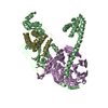
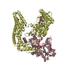
Polycomb protein ... , 2 types, 4 molecules EFST
#2: Protein
PolycombproteinEED / hEED / WD protein associating with integrin cytoplasmic tails 1 / WAIT-1
Mass: 41776.535 Da / Num. of mol.: 2 Source method: isolated from a genetically manipulated source Source: (gene. exp.) Homo sapiens (human) / Gene: EED / Production host: Spodoptera frugiperda (fall armyworm) / References: UniProt: O75530
#3: Protein
PolycombproteinSUZ12 / Chromatin precipitated E2F target 9 protein / ChET 9 protein / Joined to JAZF1 protein / Suppressor ...Chromatin precipitated E2F target 9 protein / ChET 9 protein / Joined to JAZF1 protein / Suppressor of zeste 12 protein homolog
Mass: 22268.748 Da / Num. of mol.: 2 Source method: isolated from a genetically manipulated source Source: (gene. exp.) Homo sapiens (human) / Gene: SUZ12, CHET9, JJAZ1, KIAA0160 / Production host: Spodoptera frugiperda (fall armyworm) / References: UniProt: Q15022
Protocol: SINGLE WAVELENGTH / Monochromatic (M) / Laue (L): M / Scattering type: x-ray
Radiation wavelength
Wavelength: 1 Å / Relative weight: 1
Reflection
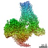
Resolution: 2.62→149.39 Å / Num. obs: 70687 / % possible obs: 99.6 % / Redundancy: 3.4 % / Biso Wilson estimate: 73.7 Å2 / Rmerge(I) obs: 0.061 / Net I/σ(I): 16.6
Reflection shell
Resolution: 2.62→3.21 Å / Redundancy: 3.4 % / Rmerge(I) obs: 0.306 / Mean I/σ(I) obs: 4.3 / % possible all: 99.9
-
Processing
Software
Name
Version
Classification
BUSTER
2.11.4
refinement
autoPROC
datascaling
Aimless
datascaling
autoSHARP
phasing
Refinement
Method to determine structure: SAD / Resolution: 2.62→91.15 Å / Cor.coef. Fo:Fc: 0.9277 / Cor.coef. Fo:Fc free: 0.8969 / SU R Cruickshank DPI: 0.701 / Cross valid method: THROUGHOUT / σ(F): 0 / SU R Blow DPI: 0.581 / SU Rfree Blow DPI: 0.282 / SU Rfree Cruickshank DPI: 0.293
Rfactor
Num. reflection
% reflection
Selection details
Rfree
0.238
3591
5.09 %
RANDOM
Rwork
0.1912
-
-
-
obs
0.1935
70532
99.6 %
-
Displacement parameters
Biso mean: 68.45 Å2
Baniso -1
Baniso -2
Baniso -3
1-
-14.6704 Å2
0 Å2
-0.3362 Å2
2-
-
18.3044 Å2
0 Å2
3-
-
-
-3.634 Å2
Refine analyze
Luzzati coordinate error obs: 0.331 Å
Refinement step
Cycle: 1 / Resolution: 2.62→91.15 Å
Protein
Nucleic acid
Ligand
Solvent
Total
Num. atoms
15526
0
86
36
15648
Refine LS restraints
Refine-ID
Type
Dev ideal
Number
Restraint function
Weight
X-RAY DIFFRACTION
t_bond_d
0.01
15954
HARMONIC
2
X-RAY DIFFRACTION
t_angle_deg
1.14
21519
HARMONIC
2
X-RAY DIFFRACTION
t_dihedral_angle_d
5716
SINUSOIDAL
2
X-RAY DIFFRACTION
t_incorr_chiral_ct
X-RAY DIFFRACTION
t_pseud_angle
X-RAY DIFFRACTION
t_trig_c_planes
448
HARMONIC
2
X-RAY DIFFRACTION
t_gen_planes
2312
HARMONIC
5
X-RAY DIFFRACTION
t_it
15954
HARMONIC
20
X-RAY DIFFRACTION
t_nbd
15
SEMIHARMONIC
5
X-RAY DIFFRACTION
t_omega_torsion
3
X-RAY DIFFRACTION
t_other_torsion
21
X-RAY DIFFRACTION
t_improper_torsion
X-RAY DIFFRACTION
t_chiral_improper_torsion
2018
SEMIHARMONIC
5
X-RAY DIFFRACTION
t_sum_occupancies
X-RAY DIFFRACTION
t_utility_distance
X-RAY DIFFRACTION
t_utility_angle
X-RAY DIFFRACTION
t_utility_torsion
X-RAY DIFFRACTION
t_ideal_dist_contact
17737
SEMIHARMONIC
4
LS refinement shell
Resolution: 2.62→2.69 Å / Total num. of bins used: 20
Rfactor
Num. reflection
% reflection
Rfree
0.2868
265
5.06 %
Rwork
0.2318
4977
-
all
0.2347
5242
-
obs
-
-
99.6 %
+
About Yorodumi
-
News
-
Feb 9, 2022. New format data for meta-information of EMDB entries
New format data for meta-information of EMDB entries
Version 3 of the EMDB header file is now the official format.
The previous official version 1.9 will be removed from the archive.
In the structure databanks used in Yorodumi, some data are registered as the other names, "COVID-19 virus" and "2019-nCoV". Here are the details of the virus and the list of structure data.
Jan 31, 2019. EMDB accession codes are about to change! (news from PDBe EMDB page)
EMDB accession codes are about to change! (news from PDBe EMDB page)
The allocation of 4 digits for EMDB accession codes will soon come to an end. Whilst these codes will remain in use, new EMDB accession codes will include an additional digit and will expand incrementally as the available range of codes is exhausted. The current 4-digit format prefixed with “EMD-” (i.e. EMD-XXXX) will advance to a 5-digit format (i.e. EMD-XXXXX), and so on. It is currently estimated that the 4-digit codes will be depleted around Spring 2019, at which point the 5-digit format will come into force.
The EM Navigator/Yorodumi systems omit the EMD- prefix.
Related info.:Q: What is EMD? / ID/Accession-code notation in Yorodumi/EM Navigator
Yorodumi is a browser for structure data from EMDB, PDB, SASBDB, etc.
This page is also the successor to EM Navigator detail page, and also detail information page/front-end page for Omokage search.
The word "yorodu" (or yorozu) is an old Japanese word meaning "ten thousand". "mi" (miru) is to see.
Related info.:EMDB / PDB / SASBDB / Comparison of 3 databanks / Yorodumi Search / Aug 31, 2016. New EM Navigator & Yorodumi / Yorodumi Papers / Jmol/JSmol / Function and homology information / Changes in new EM Navigator and Yorodumi
 Movie
Movie Controller


 Open data
 Basic information
 Components
 Keywords
 Function and homology information
 Anolis carolinensis (green anole)
 Homo sapiens (human)
 X-RAY DIFFRACTION /
  Authors
 Citation
 Structure visualization
 Downloads & links
 Other downloads

 PDBj
PDBj


 Assembly
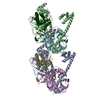


 Spodoptera frugiperda (fall armyworm)
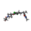


 Mass: 536.447 Da / Num. of mol.: 2 / Source method: obtained synthetically / Formula: C26H31Cl2N3O5
 Mass: 65.409 Da / Num. of mol.: 14 / Source method: obtained synthetically / Formula: Zn
 Sample preparation
 / Beamline: 17-ID / Wavelength: 1 Å
 Processing