Journal: J Struct Biol / Year: 2016 Title: The Sac3 TPR-like region in the Saccharomyces cerevisiae TREX-2 complex is more extensive but independent of the CID region. Authors: Shintaro Aibara / Xiao-Chen Bai / Murray Stewart / Abstract: Transcription-export complex 2 (TREX-2 complex) facilitates the localization of actively transcribing genes to the nuclear periphery and also functions to contribute to the generation of export- ...Transcription-export complex 2 (TREX-2 complex) facilitates the localization of actively transcribing genes to the nuclear periphery and also functions to contribute to the generation of export-competent mRNPs through interactions with the general mRNA nuclear export factor Mex67:Mtr2. The TREX-2 complex is based on a Sac3 scaffold to which Thp1, Sem1, Cdc31, and Sus1 bind. TREX-2 can be subdivided into two modules: one, in which Thp1 and Sem1 bind to the Sac3(M) region (residues ∼100-551), and the other in which Cdc31 and two Sus1 chains bind to the Sac3(CID) region (residues ∼710-805). Complementary structural analyses using X-ray crystallography, electron microscopy, and small-angle X-ray scattering of the Saccharomyces cerevisiae TREX-2 complex, expressed using Baculovirus-infected Sf9 cells, have indicated that the TPR-like repeats of the Sac3(M) region extend considerably further towards the N-terminus than previously thought, and also indicate that this region and Sac3(CID):Sus1:Cdc31 region of the S. cerevisiae complex are structurally independent. Although the density visible accounted for only ∼100kDa, a 5.3Å resolution cryo-EM reconstruction was obtained of the M-region of TREX-2 that showed an additional three putative α-helices extending towards the Sac3 N-terminus and these helices were also seen in a 4.9Å resolution structure obtained by X-ray crystallography. SUMMARY STATEMENT: We describe the expression, purification and structural characterization of the S. cerevisiae TREX-2 complex and demonstrate that the Sac3 TPR-like repeats are more extensive than ...SUMMARY STATEMENT: We describe the expression, purification and structural characterization of the S. cerevisiae TREX-2 complex and demonstrate that the Sac3 TPR-like repeats are more extensive than previously thought and that the M- and CID-regions do not appear to have a defined spatial orientation.
History
Deposition
May 26, 2016
-
Header (metadata) release
Jul 27, 2016
-
Map release
Jul 27, 2016
-
Update
Nov 23, 2016
-
Current status
Nov 23, 2016
Processing site: PDBe / Status: Released
-
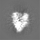
Structure visualization
Movie
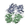
Surface view with section colored by density value
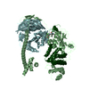
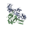
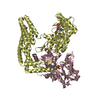
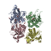
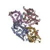
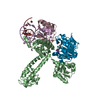
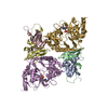
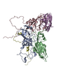
Supramolecule #1000: Sac3 in complex with Thp1 and Sem1
Supramolecule
Name: Sac3 in complex with Thp1 and Sem1 / type: sample / ID: 1000 / Details: Sample is monodisperse / Oligomeric state: One heterotrimer / Number unique components: 3
Cryogen name: ETHANE / Chamber humidity: 100 % / Chamber temperature: 85 K / Instrument: HOMEMADE PLUNGER / Method: Blot for 7 seconds before plunging from one side
-
Electron microscopy
Microscope
FEI TITAN KRIOS
Temperature
Min: 80 K / Max: 90 K / Average: 85 K
Specialist optics
Energy filter - Name: Gatan Quantum / Energy filter - Lower energy threshold: 0.0 eV / Energy filter - Upper energy threshold: 20.0 eV
Date
Nov 3, 2015
Image recording
Category: CCD / Film or detector model: GATAN K2 QUANTUM (4k x 4k) / Average electron dose: 40 e/Å2
Electron beam
Acceleration voltage: 300 kV / Electron source: FIELD EMISSION GUN
In the structure databanks used in Yorodumi, some data are registered as the other names, "COVID-19 virus" and "2019-nCoV". Here are the details of the virus and the list of structure data.
Jan 31, 2019. EMDB accession codes are about to change! (news from PDBe EMDB page)
EMDB accession codes are about to change! (news from PDBe EMDB page)
The allocation of 4 digits for EMDB accession codes will soon come to an end. Whilst these codes will remain in use, new EMDB accession codes will include an additional digit and will expand incrementally as the available range of codes is exhausted. The current 4-digit format prefixed with “EMD-” (i.e. EMD-XXXX) will advance to a 5-digit format (i.e. EMD-XXXXX), and so on. It is currently estimated that the 4-digit codes will be depleted around Spring 2019, at which point the 5-digit format will come into force.
The EM Navigator/Yorodumi systems omit the EMD- prefix.
Related info.:Q: What is EMD? / ID/Accession-code notation in Yorodumi/EM Navigator
Yorodumi is a browser for structure data from EMDB, PDB, SASBDB, etc.
This page is also the successor to EM Navigator detail page, and also detail information page/front-end page for Omokage search.
The word "yorodu" (or yorozu) is an old Japanese word meaning "ten thousand". "mi" (miru) is to see.
Related info.:EMDB / PDB / SASBDB / Comparison of 3 databanks / Yorodumi Search / Aug 31, 2016. New EM Navigator & Yorodumi / Yorodumi Papers / Jmol/JSmol / Function and homology information / Changes in new EM Navigator and Yorodumi
 Movie
Movie Controller


 Open data
 Basic information
 Map data
 Sample
 Keywords
 Function and homology information
 Authors
 Citation
 Structure visualization
 Downloads & links
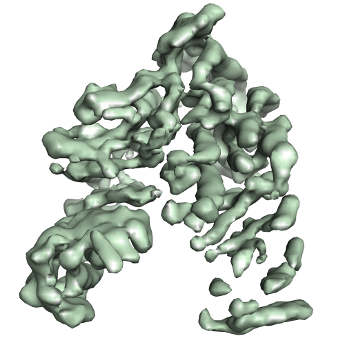 emd-3440.png
 http://ftp.pdbj.org/pub/emdb/structures/EMD-3440


 Z (Sec.)
 Y (Row.)
 X (Col.)


 Sample components
 Spodoptera frugiperda (fall armyworm) / Recombinant cell: sf9 / Recombinant plasmid: pAceBacRZ
 Processing
 Electron microscopy
 FIELD EMISSION GUN
