 Movie
Movie Controller


+ Open data

- Basic information
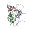
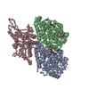
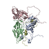
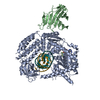
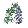
| Entry | Database: PDB / ID: 5g5p | ||||||
|---|---|---|---|---|---|---|---|
| Title | Structure of the Saccharomyces cerevisiae TREX-2 complex | ||||||
 Components Components |
| ||||||
 Keywords Keywords | TRANSPORT PROTEIN / MRNA / MRNA EXPORT | ||||||
| Function / homology |  Function and homology information Function and homology informationactin filament-based process / cellular bud site selection / SAGA complex localization to transcription regulatory region / transcription export complex 2 / nuclear pore cytoplasmic filaments / maintenance of DNA trinucleotide repeats / nuclear mRNA surveillance / post-transcriptional tethering of RNA polymerase II gene DNA at nuclear periphery / filamentous growth / mRNA 3'-end processing ...actin filament-based process / cellular bud site selection / SAGA complex localization to transcription regulatory region / transcription export complex 2 / nuclear pore cytoplasmic filaments / maintenance of DNA trinucleotide repeats / nuclear mRNA surveillance / post-transcriptional tethering of RNA polymerase II gene DNA at nuclear periphery / filamentous growth / mRNA 3'-end processing / proteasome regulatory particle, lid subcomplex / poly(A)+ mRNA export from nucleus / proteasome storage granule / proteasome assembly / mRNA export from nucleus / transcription-coupled nucleotide-excision repair / protein export from nucleus / protein folding chaperone / proteasome complex / transcription elongation by RNA polymerase II / double-strand break repair via homologous recombination / nuclear envelope / mitotic cell cycle / ribosomal small subunit biogenesis / double-stranded DNA binding / molecular adaptor activity / ubiquitin-dependent protein catabolic process / proteasome-mediated ubiquitin-dependent protein catabolic process / regulation of cell cycle / positive regulation of transcription by RNA polymerase II / RNA binding / nucleus / cytoplasm / cytosol Similarity search - Function | ||||||
| Biological species |  | ||||||
| Method | ELECTRON MICROSCOPY / single particle reconstruction / cryo EM / Resolution: 5.3 Å | ||||||
 Authors Authors | Aibara, S. / Bai, X.C. / Stewart, M. | ||||||
 Citation Citation | Journal: J Struct Biol / Year: 2016 Title: The Sac3 TPR-like region in the Saccharomyces cerevisiae TREX-2 complex is more extensive but independent of the CID region. Authors: Shintaro Aibara / Xiao-Chen Bai / Murray Stewart /  Abstract: Transcription-export complex 2 (TREX-2 complex) facilitates the localization of actively transcribing genes to the nuclear periphery and also functions to contribute to the generation of export- ...Transcription-export complex 2 (TREX-2 complex) facilitates the localization of actively transcribing genes to the nuclear periphery and also functions to contribute to the generation of export-competent mRNPs through interactions with the general mRNA nuclear export factor Mex67:Mtr2. The TREX-2 complex is based on a Sac3 scaffold to which Thp1, Sem1, Cdc31, and Sus1 bind. TREX-2 can be subdivided into two modules: one, in which Thp1 and Sem1 bind to the Sac3(M) region (residues ∼100-551), and the other in which Cdc31 and two Sus1 chains bind to the Sac3(CID) region (residues ∼710-805). Complementary structural analyses using X-ray crystallography, electron microscopy, and small-angle X-ray scattering of the Saccharomyces cerevisiae TREX-2 complex, expressed using Baculovirus-infected Sf9 cells, have indicated that the TPR-like repeats of the Sac3(M) region extend considerably further towards the N-terminus than previously thought, and also indicate that this region and Sac3(CID):Sus1:Cdc31 region of the S. cerevisiae complex are structurally independent. Although the density visible accounted for only ∼100kDa, a 5.3Å resolution cryo-EM reconstruction was obtained of the M-region of TREX-2 that showed an additional three putative α-helices extending towards the Sac3 N-terminus and these helices were also seen in a 4.9Å resolution structure obtained by X-ray crystallography. SUMMARY STATEMENT: We describe the expression, purification and structural characterization of the S. cerevisiae TREX-2 complex and demonstrate that the Sac3 TPR-like repeats are more extensive than ...SUMMARY STATEMENT: We describe the expression, purification and structural characterization of the S. cerevisiae TREX-2 complex and demonstrate that the Sac3 TPR-like repeats are more extensive than previously thought and that the M- and CID-regions do not appear to have a defined spatial orientation. | ||||||
| History |
|
- Structure visualization
| Movie |
Movie viewer |
|---|---|
| Structure viewer | Molecule: MolmilJmol/JSmol |
- Downloads & links
-Download
| PDBx/mmCIF format | 5g5p.cif.gz | 220.5 KB | Display | PDBx/mmCIF format |
|---|---|---|---|---|
| PDB format | pdb5g5p.ent.gz | 150.1 KB | Display | PDB format |
| PDBx/mmJSON format | 5g5p.json.gz | Tree view | PDBx/mmJSON format | |
| Others |  Other downloads Other downloads |
-Validation report
| Arichive directory | https://data.pdbj.org/pub/pdb/validation_reports/g5/5g5pftp://data.pdbj.org/pub/pdb/validation_reports/g5/5g5p | HTTPS FTP |
|---|
-Related structure data
| Related structure data | 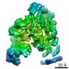 3440MC  5l3tC M: map data used to model this data C: citing same article ( |
|---|---|
| Similar structure data |


-Links
 PDBj
PDBj

- Assembly
| Deposited unit | 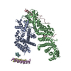
|
|---|---|
| 1 |
|
-Components
| #1: Protein | Mass: 93213.320 Da / Num. of mol.: 1 Source method: isolated from a genetically manipulated source Source: (gene. exp.) Plasmid: PACEBACRZ / Cell line (production host): SF9 / Production host:   SPODOPTERA FRUGIPERDA (fall armyworm) / References: UniProt: P46674 SPODOPTERA FRUGIPERDA (fall armyworm) / References: UniProt: P46674 | ||
|---|---|---|---|
| #2: Protein | Mass: 52734.820 Da / Num. of mol.: 1 Source method: isolated from a genetically manipulated source Source: (gene. exp.) Plasmid: PACEBACRZ / Cell line (production host): SF9 / Production host: SPODOPTERA FRUGIPERDA (fall armyworm) / References: UniProt: Q08231 | ||
| #3: Protein | Mass: 10397.102 Da / Num. of mol.: 1 Source method: isolated from a genetically manipulated source Source: (gene. exp.) Plasmid: PACEBACRZ / Cell line (production host): SF9 / Production host: SPODOPTERA FRUGIPERDA (fall armyworm) / References: UniProt: O94742 | ||
| #4: Protein | Mass: 68527.133 Da / Num. of mol.: 3 Source method: isolated from a genetically manipulated source Details: CHAINS D, F AND E ARE MODELLED AS POLYALA Source: (gene. exp.) Plasmid: PACEBACRZ / Cell line (production host): SF9 / Production host: SPODOPTERA FRUGIPERDA (fall armyworm)Sequence details | CHAINS D, E AND F ARE BUILT AS POLYALA DUE TO RESOLUTION | |
-Experimental details
-Experiment
| Experiment | Method: ELECTRON MICROSCOPY |
|---|---|
| EM experiment | Aggregation state: PARTICLE / 3D reconstruction method: single particle reconstruction |
- Sample preparation
| Component | Name: Sac3 in complex with Thp1 and Sem1 / Type: COMPLEX / Oligomeric details: One heterotrimer / Source: MULTIPLE SOURCES |
|---|---|
| Molecular weight | Value: 0.19 MDa / Experimental value: NO |
| Buffer solution | Name: 20 MM HEPES, 300 MM NACL, 5 MM DTT / pH: 8 / Details: 20 mM HEPES, 300 mM NaCl, 5 mM DTT |
| Specimen | Conc.: 0.015 mg/ml / Embedding applied: NO / Shadowing applied: NO / Staining applied: NO / Vitrification applied: YES |
| Specimen support | Details: OTHER |
| Vitrification | Instrument: HOMEMADE PLUNGER / Cryogen name: ETHANE / Temp: 85 K / Humidity: 100 % / Details: LIQUID ETHANE |
- Electron microscopy imaging
| Experimental equipment |  Model: Titan Krios / Image courtesy: FEI Company |
|---|---|
| Microscopy | Model: FEI TITAN KRIOS / Date: Nov 3, 2015 |
| Electron gun | Electron source:  FIELD EMISSION GUN / Accelerating voltage: 300 kV / Illumination mode: FLOOD BEAM FIELD EMISSION GUN / Accelerating voltage: 300 kV / Illumination mode: FLOOD BEAM |
| Electron lens | Mode: BRIGHT FIELD / Nominal magnification: 81000 X / Calibrated magnification: 35714 X / Nominal defocus max: 4500 nm / Nominal defocus min: 800 nm / Cs: 2.7 mm |
| Specimen holder | Temperature: 85 K / Temperature (max): 90 K / Temperature (min): 80 K |
| Image recording | Electron dose: 40 e/Å2 / Film or detector model: GATAN K2 QUANTUM (4k x 4k) |
| Image scans | Num. digital images: 496 |
- Processing
| EM software | Name: RELION / Category: 3D reconstruction | ||||||||||||
|---|---|---|---|---|---|---|---|---|---|---|---|---|---|
| Particle selection | Details: The particles were processed using Relion | ||||||||||||
| Symmetry | Point symmetry: C1 (asymmetric) | ||||||||||||
| 3D reconstruction | Resolution: 5.3 Å / Resolution method: FSC 0.143 CUT-OFF / Num. of particles: 81559 / Refinement type: HALF-MAPS REFINED INDEPENDENTLY / Symmetry type: POINT | ||||||||||||
| Refinement | Highest resolution: 5.3 Å | ||||||||||||
| Refinement step | Cycle: LAST / Highest resolution: 5.3 Å
|
