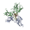
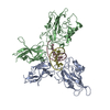
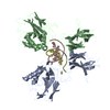
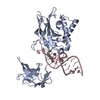
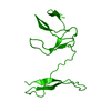
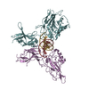
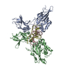
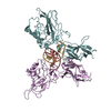
Entry Database : PDB / ID : 2v2tTitle X-ray structure of a NF-kB p50-RelB-DNA complex 5'-D(*CP*GP*GP*GP*AP*AP*TP*TP*CP*CP*CP)-3'NUCLEAR FACTOR NF-KAPPA-B P105 SUBUNIT TRANSCRIPTION FACTOR RELB Keywords / / / / / / / / Function / homology Function Domain/homology Component
/ / / / / / / / / / / / / / / / / / / / / / / / / / / / / / / / / / / / / / / / / / / / / / / / / / / / / / / / / / / / / / / / / / / / / / / / / / / / / / / / / / / / / / / / / / / / / / / / / / / / / / / / / / / / / / / / / / / / / / / / / / / / / / / / / / / / / / / / / / / / / / / / / / / / / / / / / / Biological species MUS MUSCULUS (house mouse)Method / / / Resolution : 3.05 Å Authors Moorthy, A.K. / Huang, D.B. / Wang, V.Y. / Vu, D. / Ghosh, G. Journal : J.Mol.Biol. / Year : 2007Title : X-Ray Structure of a NF-kappaB p50/Relb/DNA Complex Reveals Assembly of Multiple Dimers on Tandem kappaB Sites.Authors : Moorthy, A.K. / Huang, D.B. / Wang, V.Y. / Vu, D. / Ghosh, G. History Deposition Jun 7, 2007 Deposition site / Processing site Revision 1.0 Jul 3, 2007 Provider / Type Revision 1.1 May 8, 2011 Group Revision 1.2 Jul 13, 2011 Group Revision 1.3 Dec 13, 2023 Group / Database references / Refinement descriptionCategory chem_comp_atom / chem_comp_bond ... chem_comp_atom / chem_comp_bond / database_2 / pdbx_initial_refinement_model Item / _database_2.pdbx_database_accessionRevision 1.4 Nov 6, 2024 Group / Category / pdbx_modification_feature
Show all Show less Remark 700 SHEET THE SHEET STRUCTURE OF THIS MOLECULE IS BIFURCATED. IN ORDER TO REPRESENT THIS FEATURE IN ... SHEET THE SHEET STRUCTURE OF THIS MOLECULE IS BIFURCATED. IN ORDER TO REPRESENT THIS FEATURE IN THE SHEET RECORDS BELOW, TWO SHEETS ARE DEFINED.
 Movie
Movie Controller


 Open data
 Basic information
 Components
 Keywords
 Function and homology information
 X-RAY DIFFRACTION /
  Authors
 Citation
 Structure visualization
 Downloads & links
 Other downloads


 PDBj
PDBj


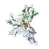
 Assembly

 Mass: 18.015 Da / Num. of mol.: 34 / Source method: isolated from a natural source / Formula: H2O
 Sample preparation
 / Beamline: 19-ID / Wavelength: 1.05
  Processing