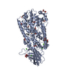
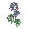
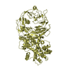
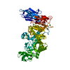
 Movie
Movie Controller


+ Open data

- Basic information
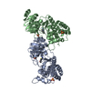
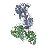
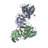
| Entry | Database: PDB / ID: 1ikn | ||||||
|---|---|---|---|---|---|---|---|
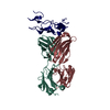
| Title | IKAPPABALPHA/NF-KAPPAB COMPLEX | ||||||
 Components Components |
| ||||||
 Keywords Keywords | TRANSCRIPTION FACTOR / IKB-NFKB COMPLEX | ||||||
| Function / homology |  Function and homology information Function and homology informationnegative regulation of cholesterol transport / cellular response to diterpene / cellular response to glucoside / SUMOylation of immune response proteins / Regulated proteolysis of p75NTR / I-kappaB/NF-kappaB complex / negative regulation of vitamin D biosynthetic process / DEx/H-box helicases activate type I IFN and inflammatory cytokines production / Interleukin-1 processing / RIP-mediated NFkB activation via ZBP1 ...negative regulation of cholesterol transport / cellular response to diterpene / cellular response to glucoside / SUMOylation of immune response proteins / Regulated proteolysis of p75NTR / I-kappaB/NF-kappaB complex / negative regulation of vitamin D biosynthetic process / DEx/H-box helicases activate type I IFN and inflammatory cytokines production / Interleukin-1 processing / RIP-mediated NFkB activation via ZBP1 / TRAF6 mediated NF-kB activation / positive regulation of hyaluronan biosynthetic process / positive regulation of Schwann cell differentiation / PKMTs methylate histone lysines / MAP3K8 (TPL2)-dependent MAPK1/3 activation / NF-kB is activated and signals survival / mammary gland involution / Turbulent (oscillatory, disturbed) flow shear stress activates signaling by PIEZO1 and integrins in endothelial cells / cellular response to carbohydrate stimulus / TAK1-dependent IKK and NF-kappa-B activation / Activation of NF-kappaB in B cells / prolactin signaling pathway / nucleotide-binding oligomerization domain containing 1 signaling pathway / positive regulation of chondrocyte differentiation / negative regulation of myeloid cell differentiation / cellular response to interleukin-17 / NF-kappaB p50/p65 complex / IkBA variant leads to EDA-ID / toll-like receptor TLR6:TLR2 signaling pathway / CLEC7A (Dectin-1) signaling / FCERI mediated NF-kB activation / Interleukin-1 signaling / antibacterial innate immune response / Downstream TCR signaling / CD209 (DC-SIGN) signaling / cellular response to peptide / negative regulation of interleukin-12 production / cellular response to peptidoglycan / macrophage activation / positive regulation of lipid storage / nucleotide-binding oligomerization domain containing 2 signaling pathway / SUMOylation of immune response proteins / ankyrin repeat binding / RIP-mediated NFkB activation via ZBP1 / negative regulation of protein sumoylation / postsynapse to nucleus signaling pathway / defense response to tumor cell / signal transduction involved in regulation of gene expression / positive regulation of T cell receptor signaling pathway / cellular response to interleukin-6 / positive regulation of macrophage derived foam cell differentiation / negative regulation of non-canonical NF-kappaB signal transduction / actinin binding / cellular response to dsRNA / cellular response to hepatocyte growth factor stimulus / positive regulation of miRNA metabolic process / : / interleukin-1-mediated signaling pathway / response to UV-B / positive regulation of leukocyte adhesion to vascular endothelial cell / nuclear localization sequence binding / toll-like receptor 4 signaling pathway / negative regulation of protein import into nucleus / negative regulation of cytokine production / positive regulation of amyloid-beta formation / vascular endothelial growth factor signaling pathway / non-canonical NF-kappaB signal transduction / NF-kappaB complex / cellular response to cold / cellular response to lipoteichoic acid / response to exogenous dsRNA / phosphate ion binding / cellular response to cytokine stimulus / response to muramyl dipeptide / hair follicle development / TRAF6 mediated NF-kB activation / cellular response to angiotensin / negative regulation of Notch signaling pathway / negative regulation of macrophage derived foam cell differentiation / negative regulation of lipid storage / positive regulation of cholesterol efflux / positive regulation of vascular endothelial growth factor production / molecular sequestering activity / lymph node development / cellular response to interleukin-1 / general transcription initiation factor binding / positive regulation of transcription initiation by RNA polymerase II / Notch signaling pathway / NF-kappaB binding / RNA polymerase II core promoter sequence-specific DNA binding / transcription regulator inhibitor activity / cis-regulatory region sequence-specific DNA binding / JNK cascade / positive regulation of interleukin-12 production / canonical NF-kappaB signal transduction / response to muscle stretch / cellular response to brain-derived neurotrophic factor stimulus / heat shock protein binding / Neutrophil degranulation / peptide binding Similarity search - Function | ||||||
| Biological species |   Homo sapiens (human) Homo sapiens (human) | ||||||
| Method |  X-RAY DIFFRACTION / SYNCHROTRON / MOLECULAR REPLACEMENT / Resolution: 2.3 Å X-RAY DIFFRACTION / SYNCHROTRON / MOLECULAR REPLACEMENT / Resolution: 2.3 Å | ||||||
 Authors Authors | Huxford, T. / Huang, D.-B. / Malek, S. / Ghosh, G. | ||||||
 Citation Citation | Journal: Cell(Cambridge,Mass.) / Year: 1998 Title: The crystal structure of the IkappaBalpha/NF-kappaB complex reveals mechanisms of NF-kappaB inactivation. Authors: Huxford, T. / Huang, D.B. / Malek, S. / Ghosh, G. | ||||||
| History |
|
- Structure visualization
| Structure viewer | Molecule: MolmilJmol/JSmol |
|---|
- Downloads & links
-Download
| PDBx/mmCIF format | 1ikn.cif.gz | 135.9 KB | Display | PDBx/mmCIF format |
|---|---|---|---|---|
| PDB format | pdb1ikn.ent.gz | 104.4 KB | Display | PDB format |
| PDBx/mmJSON format | 1ikn.json.gz | Tree view | PDBx/mmJSON format | |
| Others |  Other downloads Other downloads |
-Validation report
| Arichive directory | https://data.pdbj.org/pub/pdb/validation_reports/ik/1iknftp://data.pdbj.org/pub/pdb/validation_reports/ik/1ikn | HTTPS FTP |
|---|
-Related structure data
| Related structure data | 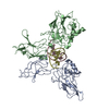 1vkxS S: Starting model for refinement |
|---|---|
| Similar structure data |


-Links
 PDBj
PDBj


- Assembly
| Deposited unit | 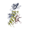
| ||||||||
|---|---|---|---|---|---|---|---|---|---|
| 1 |
| ||||||||
| Unit cell |
|
-Components
| #1: Protein | Mass: 32738.027 Da / Num. of mol.: 1 / Fragment: N-TERMINAL AND DIMERIZATION DOMAINS Source method: isolated from a genetically manipulated source Source: (gene. exp.)  |
|---|---|
| #2: Protein | Mass: 13929.799 Da / Num. of mol.: 1 / Fragment: N-TERMINAL AND DIMERIZATION DOMAINS Source method: isolated from a genetically manipulated source Source: (gene. exp.) |
| #3: Protein | Mass: 26098.195 Da / Num. of mol.: 1 Source method: isolated from a genetically manipulated source Source: (gene. exp.) Homo sapiens (human) / Gene: MAD-3 / Species (production host): Escherichia coli / Production host: |
| #4: Water | ChemComp-HOH /  Mass: 18.015 Da / Num. of mol.: 212 / Source method: isolated from a natural source / Formula: H2O Mass: 18.015 Da / Num. of mol.: 212 / Source method: isolated from a natural source / Formula: H2O |
-Experimental details
-Experiment
| Experiment | Method: X-RAY DIFFRACTION / Number of used crystals: 5 |
|---|
- Sample preparation
| Crystal | Density Matthews: 2.41 Å3/Da / Density % sol: 43 % | ||||||||||||||||||||||||||||||||||||||||
|---|---|---|---|---|---|---|---|---|---|---|---|---|---|---|---|---|---|---|---|---|---|---|---|---|---|---|---|---|---|---|---|---|---|---|---|---|---|---|---|---|---|
| Crystal grow | pH: 7 / Details: pH 7.00 | ||||||||||||||||||||||||||||||||||||||||
| Crystal | *PLUS Density % sol: 43 % | ||||||||||||||||||||||||||||||||||||||||
| Crystal grow | *PLUS Temperature: 23-24 ℃ / Method: vapor diffusion, hanging drop | ||||||||||||||||||||||||||||||||||||||||
| Components of the solutions | *PLUS
|
-Data collection
| Diffraction | Mean temperature: 105 K |
|---|---|
| Diffraction source | Source: SYNCHROTRON / Site: NSLS  / Beamline: X25 / Wavelength: 1.54 / Beamline: X25 / Wavelength: 1.54 |
| Detector | Detector: CCD |
| Radiation | Protocol: SINGLE WAVELENGTH / Monochromatic (M) / Laue (L): M / Scattering type: x-ray |
| Radiation wavelength | Wavelength: 1.54 Å / Relative weight: 1 |
| Reflection | Resolution: 2.3→30 Å / Num. obs: 28014 / % possible obs: 91 % / Observed criterion σ(I): 1 / Redundancy: 3.5 % / Biso Wilson estimate: 25.5 Å2 / Rmerge(I) obs: 0.043 / Net I/σ(I): 15.1 |
| Reflection shell | Resolution: 2.3→2.38 Å / Rmerge(I) obs: 0.22 / Mean I/σ(I) obs: 2.5 / % possible all: 59 |
| Reflection | *PLUS Highest resolution: 2.3 Å / Lowest resolution: 30 Å / % possible obs: 91 % / Redundancy: 3.5 % / Num. measured all: 160828 |
| Reflection shell | *PLUS Highest resolution: 2.3 Å / Lowest resolution: 2.38 Å / % possible obs: 59 % / Mean I/σ(I) obs: 2.5 |
- Processing
| Software |
| ||||||||||||||||||||||||||||||||||||||||||||||||||||||||||||
|---|---|---|---|---|---|---|---|---|---|---|---|---|---|---|---|---|---|---|---|---|---|---|---|---|---|---|---|---|---|---|---|---|---|---|---|---|---|---|---|---|---|---|---|---|---|---|---|---|---|---|---|---|---|---|---|---|---|---|---|---|---|
| Refinement | Method to determine structure: MOLECULAR REPLACEMENT Starting model: PDB ENTRY 1VKX Resolution: 2.3→6 Å / σ(F): 3
| ||||||||||||||||||||||||||||||||||||||||||||||||||||||||||||
| Displacement parameters | Biso mean: 48.6 Å2
| ||||||||||||||||||||||||||||||||||||||||||||||||||||||||||||
| Refine analyze |
| ||||||||||||||||||||||||||||||||||||||||||||||||||||||||||||
| Refinement step | Cycle: LAST / Resolution: 2.3→6 Å
| ||||||||||||||||||||||||||||||||||||||||||||||||||||||||||||
| Refine LS restraints |
| ||||||||||||||||||||||||||||||||||||||||||||||||||||||||||||
| LS refinement shell | Resolution: 2.3→2.44 Å / Total num. of bins used: 6
| ||||||||||||||||||||||||||||||||||||||||||||||||||||||||||||
| Xplor file |
| ||||||||||||||||||||||||||||||||||||||||||||||||||||||||||||
| Software | *PLUS Name: CNS / Version: 0.4 / Classification: refinement | ||||||||||||||||||||||||||||||||||||||||||||||||||||||||||||
| Refinement | *PLUS Rfactor obs: 0.223 | ||||||||||||||||||||||||||||||||||||||||||||||||||||||||||||
| Solvent computation | *PLUS | ||||||||||||||||||||||||||||||||||||||||||||||||||||||||||||
| Displacement parameters | *PLUS | ||||||||||||||||||||||||||||||||||||||||||||||||||||||||||||
| Refine LS restraints | *PLUS
| ||||||||||||||||||||||||||||||||||||||||||||||||||||||||||||
| LS refinement shell | *PLUS Rfactor obs: 0.3 |
