 Movie
Movie Controller

[English] 日本語
 Yorodumi
- PDB-4c29: Crystal Structure of High-Affinity von Willebrand Factor A1 domai... -
+ Open data

- Basic information
| Entry | Database: PDB / ID: 4c29 | ||||||
|---|---|---|---|---|---|---|---|
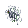
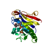
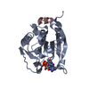
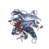
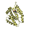
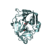
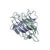
| Title | Crystal Structure of High-Affinity von Willebrand Factor A1 domain with Disulfide Mutation | ||||||
 Components Components | VON WILLEBRAND FACTOR | ||||||
 Keywords Keywords | BLOOD CLOTTING / CELL ADHESION | ||||||
| Function / homology |  Function and homology information Function and homology informationDefective VWF binding to collagen type I / Enhanced cleavage of VWF variant by ADAMTS13 / Defective VWF cleavage by ADAMTS13 variant / Defective F8 binding to von Willebrand factor / Enhanced binding of GP1BA variant to VWF multimer:collagen / Defective binding of VWF variant to GPIb:IX:V / Weibel-Palade body / hemostasis / platelet alpha granule / Platelet Adhesion to exposed collagen ...Defective VWF binding to collagen type I / Enhanced cleavage of VWF variant by ADAMTS13 / Defective VWF cleavage by ADAMTS13 variant / Defective F8 binding to von Willebrand factor / Enhanced binding of GP1BA variant to VWF multimer:collagen / Defective binding of VWF variant to GPIb:IX:V / Weibel-Palade body / hemostasis / platelet alpha granule / Platelet Adhesion to exposed collagen / extracellular matrix structural constituent / GP1b-IX-V activation signalling / p130Cas linkage to MAPK signaling for integrins / Defective F8 cleavage by thrombin / Platelet Aggregation (Plug Formation) / cell-substrate adhesion / GRB2:SOS provides linkage to MAPK signaling for Integrins / positive regulation of intracellular signal transduction / immunoglobulin binding / Integrin cell surface interactions / collagen binding / : / Integrin signaling / platelet alpha granule lumen / Signaling by high-kinase activity BRAF mutants / platelet activation / MAP2K and MAPK activation / response to wounding / integrin binding / blood coagulation / Signaling by RAF1 mutants / Signaling by moderate kinase activity BRAF mutants / Paradoxical activation of RAF signaling by kinase inactive BRAF / Signaling downstream of RAS mutants / Signaling by BRAF and RAF1 fusions / Platelet degranulation / protein-folding chaperone binding / extracellular matrix / protease binding / cell adhesion / endoplasmic reticulum / : / extracellular exosome / extracellular region / identical protein binding Similarity search - Function | ||||||
| Biological species |  HOMO SAPIENS (human) HOMO SAPIENS (human) | ||||||
| Method |  X-RAY DIFFRACTION / SYNCHROTRON / MOLECULAR REPLACEMENT / Resolution: 2.202 Å X-RAY DIFFRACTION / SYNCHROTRON / MOLECULAR REPLACEMENT / Resolution: 2.202 Å | ||||||
 Authors Authors | Blenner, M.A. / Dong, X. / Springer, T.A. | ||||||
 Citation Citation | Journal: J.Biol.Chem. / Year: 2014 Title: Towards the Structural Basis of Regulation of Von Willebrand Factor Binding to Glycoprotein Ib Authors: Blenner, M.A. / Dong, X. / Springer, T.A. | ||||||
| History |
|
- Structure visualization
| Structure viewer | Molecule: MolmilJmol/JSmol |
|---|
- Downloads & links
-Download
| PDBx/mmCIF format | 4c29.cif.gz | 182.2 KB | Display | PDBx/mmCIF format |
|---|---|---|---|---|
| PDB format | pdb4c29.ent.gz | 145.7 KB | Display | PDB format |
| PDBx/mmJSON format | 4c29.json.gz | Tree view | PDBx/mmJSON format | |
| Others |  Other downloads Other downloads |
-Validation report
| Arichive directory | https://data.pdbj.org/pub/pdb/validation_reports/c2/4c29ftp://data.pdbj.org/pub/pdb/validation_reports/c2/4c29 | HTTPS FTP |
|---|
-Related structure data
| Related structure data | 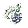 4c2aC 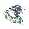 4c2bC  1auqS C: citing same article ( S: Starting model for refinement |
|---|---|
| Similar structure data |


-Links
 PDBj
PDBj


- Assembly
| Deposited unit | 
| ||||||||
|---|---|---|---|---|---|---|---|---|---|
| 1 | 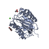
| ||||||||
| 2 | 
| ||||||||
| Unit cell |
|
-Components
| #1: Protein | Mass: 24706.678 Da / Num. of mol.: 2 / Fragment: RESIDUES 1264-1471 / Mutation: YES Source method: isolated from a genetically manipulated source Details: HIGH AFFINITY MUTANT OF A1 / Source: (gene. exp.) HOMO SAPIENS (human) / Production host:  #2: Chemical | ChemComp-ACT / |   Mass: 59.044 Da / Num. of mol.: 1 / Source method: obtained synthetically / Formula: C2H3O2 Mass: 59.044 Da / Num. of mol.: 1 / Source method: obtained synthetically / Formula: C2H3O2#3: Chemical | ChemComp-PEG /   Mass: 106.120 Da / Num. of mol.: 4 / Source method: obtained synthetically / Formula: C4H10O3 Mass: 106.120 Da / Num. of mol.: 4 / Source method: obtained synthetically / Formula: C4H10O3#4: Chemical |   Mass: 40.078 Da / Num. of mol.: 3 / Source method: obtained synthetically / Formula: Ca Mass: 40.078 Da / Num. of mol.: 3 / Source method: obtained synthetically / Formula: Ca#5: Water | ChemComp-HOH / |  Mass: 18.015 Da / Num. of mol.: 175 / Source method: isolated from a natural source / Formula: H2O Mass: 18.015 Da / Num. of mol.: 175 / Source method: isolated from a natural source / Formula: H2OHas protein modification | Y | |
|---|
-Experimental details
-Experiment
| Experiment | Method: X-RAY DIFFRACTION / Number of used crystals: 1 |
|---|
- Sample preparation
| Crystal | Density Matthews: 2.06 Å3/Da / Density % sol: 40.2 % / Description: NONE |
|---|---|
| Crystal grow | pH: 4.6 Details: CRYSTALS OF A1/SS APPEARED IN DROPS CONTAINING 25% PEG3350 AND 0.2 M CALCIUM ACETATE. THESE CRYSTALS WERE CRUSHED AND USED FOR SEEDING. CRYSTALS GROWN IN 8 MG/ML PROTEIN, 15% PEG3350, AND 0. ...Details: CRYSTALS OF A1/SS APPEARED IN DROPS CONTAINING 25% PEG3350 AND 0.2 M CALCIUM ACETATE. THESE CRYSTALS WERE CRUSHED AND USED FOR SEEDING. CRYSTALS GROWN IN 8 MG/ML PROTEIN, 15% PEG3350, AND 0.2 M CALCIUM ACETATE WERE HARVESTED, AND SOAKED IN THE SAME BUFFER CONTAINING 20% GLYCEROL FOR CRYOPROTECTION., pH 4.6 |
-Data collection
| Diffraction | Mean temperature: 93 K |
|---|---|
| Diffraction source | Source: SYNCHROTRON / Site: APS  / Beamline: 23-ID / Wavelength: 1.03322 / Beamline: 23-ID / Wavelength: 1.03322 |
| Detector | Type: MARRESEARCH / Detector: CCD / Date: Apr 5, 2012 |
| Radiation | Protocol: SINGLE WAVELENGTH / Monochromatic (M) / Laue (L): M / Scattering type: x-ray |
| Radiation wavelength | Wavelength: 1.03322 Å / Relative weight: 1 |
| Reflection | Resolution: 2.2→50 Å / Num. obs: 20133 / % possible obs: 99.5 % / Observed criterion σ(I): 2 / Redundancy: 2.5 % / Biso Wilson estimate: 29.76 Å2 / Rmerge(I) obs: 0.15 / Net I/σ(I): 13.2 |
| Reflection shell | Resolution: 2.2→2.24 Å / Rmerge(I) obs: 0.54 / Mean I/σ(I) obs: 2 / % possible all: 98 |
- Processing
| Software |
| ||||||||||||||||||||||||||||||||||||||||||||||||||||||||
|---|---|---|---|---|---|---|---|---|---|---|---|---|---|---|---|---|---|---|---|---|---|---|---|---|---|---|---|---|---|---|---|---|---|---|---|---|---|---|---|---|---|---|---|---|---|---|---|---|---|---|---|---|---|---|---|---|---|
| Refinement | Method to determine structure: MOLECULAR REPLACEMENT Starting model: PDB ENTRY 1AUQ Resolution: 2.202→41.202 Å / SU ML: 0.26 / σ(F): 1.34 / Phase error: 26.27 / Stereochemistry target values: ML
| ||||||||||||||||||||||||||||||||||||||||||||||||||||||||
| Solvent computation | Shrinkage radii: 0.9 Å / VDW probe radii: 1.11 Å / Solvent model: FLAT BULK SOLVENT MODEL | ||||||||||||||||||||||||||||||||||||||||||||||||||||||||
| Refinement step | Cycle: LAST / Resolution: 2.202→41.202 Å
| ||||||||||||||||||||||||||||||||||||||||||||||||||||||||
| Refine LS restraints |
| ||||||||||||||||||||||||||||||||||||||||||||||||||||||||
| LS refinement shell |
| ||||||||||||||||||||||||||||||||||||||||||||||||||||||||
| Refinement TLS params. | Method: refined / Origin x: 7.148 Å / Origin y: 6.7977 Å / Origin z: -23.7497 Å
| ||||||||||||||||||||||||||||||||||||||||||||||||||||||||
| Refinement TLS group | Selection details: ALL |
