- PDB-3hxg: Crystal structure of Schistsome eIF4E complexed with m7GpppA and 4E-BP -
+
Open data
ID or keywords:
Loading...
-
Basic information
Entry
Database: PDB / ID: 3hxg
Title
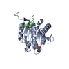
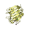
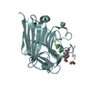
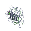
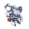
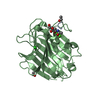
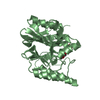
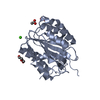
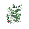
Crystal structure of Schistsome eIF4E complexed with m7GpppA and 4E-BP
Components
Eukaryotic Translation Initiation Factor 4E
Eukaryotic translation initiation factor 4E-binding protein 1
Keywords
TRANSLATION / protein-mRNA cap complex / Acetylation / Phosphoprotein / Protein synthesis inhibitor / Translation regulation
Function / homology
Function and homology information
Activation of the mRNA upon binding of the cap-binding complex and eIFs, and subsequent binding to 43S / eukaryotic initiation factor 4E binding / TOR signaling / mTORC1-mediated signalling / translation initiation factor binding / translation repressor activity / positive regulation of mitotic cell cycle / negative regulation of translational initiation / G1/S transition of mitotic cell cycle / negative regulation of translation ...Activation of the mRNA upon binding of the cap-binding complex and eIFs, and subsequent binding to 43S / eukaryotic initiation factor 4E binding / TOR signaling / mTORC1-mediated signalling / translation initiation factor binding / translation repressor activity / positive regulation of mitotic cell cycle / negative regulation of translational initiation / G1/S transition of mitotic cell cycle / negative regulation of translation / nucleus / cytosol Similarity search - Function
Mass: 22237.830 Da / Num. of mol.: 1 Source method: isolated from a genetically manipulated source Source: (gene. exp.) Schistosoma mansoni (invertebrata) / Gene: eif4e / Plasmid: pGEX-6p-1 / Production host: Escherichia coli (E. coli) / Strain (production host): XA90
#2: Protein/peptide
Eukaryotictranslationinitiationfactor4E-bindingprotein1 / eIF4E-binding protein 1 / 4E-BP1 / Phosphorylated heat- and acid-stable protein regulated by ...eIF4E-binding protein 1 / 4E-BP1 / Phosphorylated heat- and acid-stable protein regulated by insulin 1 / PHAS-I
Mass: 2432.823 Da / Num. of mol.: 1 / Fragment: residues 51-67 / Source method: obtained synthetically / Source: (synth.) Homo sapiens (human) / References: UniProt: Q13541
In the structure databanks used in Yorodumi, some data are registered as the other names, "COVID-19 virus" and "2019-nCoV". Here are the details of the virus and the list of structure data.
Jan 31, 2019. EMDB accession codes are about to change! (news from PDBe EMDB page)
EMDB accession codes are about to change! (news from PDBe EMDB page)
The allocation of 4 digits for EMDB accession codes will soon come to an end. Whilst these codes will remain in use, new EMDB accession codes will include an additional digit and will expand incrementally as the available range of codes is exhausted. The current 4-digit format prefixed with “EMD-” (i.e. EMD-XXXX) will advance to a 5-digit format (i.e. EMD-XXXXX), and so on. It is currently estimated that the 4-digit codes will be depleted around Spring 2019, at which point the 5-digit format will come into force.
The EM Navigator/Yorodumi systems omit the EMD- prefix.
Related info.:Q: What is EMD? / ID/Accession-code notation in Yorodumi/EM Navigator
Yorodumi is a browser for structure data from EMDB, PDB, SASBDB, etc.
This page is also the successor to EM Navigator detail page, and also detail information page/front-end page for Omokage search.
The word "yorodu" (or yorozu) is an old Japanese word meaning "ten thousand". "mi" (miru) is to see.
Related info.:EMDB / PDB / SASBDB / Comparison of 3 databanks / Yorodumi Search / Aug 31, 2016. New EM Navigator & Yorodumi / Yorodumi Papers / Jmol/JSmol / Function and homology information / Changes in new EM Navigator and Yorodumi
 Movie
Movie Controller

 Yorodumi
 Open data
 Basic information
 Components
 Keywords
 Function and homology information
 Homo sapiens (human)
 X-RAY DIFFRACTION /
  Authors
 Citation
 Structure visualization
 Downloads & links
 Other downloads


 PDBj
PDBj
 Assembly
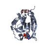

 Mass: 787.441 Da / Num. of mol.: 1 / Source method: obtained synthetically / Formula: C21H30N10O17P3
 Mass: 18.015 Da / Num. of mol.: 114 / Source method: isolated from a natural source / Formula: H2O
 Sample preparation
 / Beamline: 4.2.2 / Wavelength: 1.5 Å
 Processing