 Movie
Movie Controller

[English] 日本語
 Yorodumi
- PDB-4c2b: Crystal Structure of High-Affinity von Willebrand Factor A1 domai... -
+ Open data

- Basic information
| Entry | Database: PDB / ID: 4c2b | ||||||
|---|---|---|---|---|---|---|---|
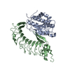
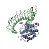
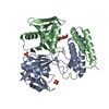
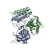
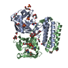
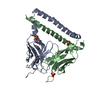
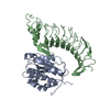
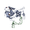
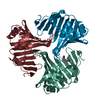
| Title | Crystal Structure of High-Affinity von Willebrand Factor A1 domain with Disulfide Mutation in Complex with High Affinity GPIb alpha | ||||||
 Components Components |
| ||||||
 Keywords Keywords | BLOOD CLOTTING | ||||||
| Function / homology |  Function and homology information Function and homology informationthrombin-activated receptor activity / glycoprotein Ib-IX-V complex / Defective VWF binding to collagen type I / Enhanced cleavage of VWF variant by ADAMTS13 / Defective VWF cleavage by ADAMTS13 variant / Defective F8 binding to von Willebrand factor / Enhanced binding of GP1BA variant to VWF multimer:collagen / Defective binding of VWF variant to GPIb:IX:V / Weibel-Palade body / blood coagulation, intrinsic pathway ...thrombin-activated receptor activity / glycoprotein Ib-IX-V complex / Defective VWF binding to collagen type I / Enhanced cleavage of VWF variant by ADAMTS13 / Defective VWF cleavage by ADAMTS13 variant / Defective F8 binding to von Willebrand factor / Enhanced binding of GP1BA variant to VWF multimer:collagen / Defective binding of VWF variant to GPIb:IX:V / Weibel-Palade body / blood coagulation, intrinsic pathway / hemostasis / Defective F9 activation / platelet alpha granule / Platelet Adhesion to exposed collagen / positive regulation of platelet activation / megakaryocyte development / extracellular matrix structural constituent / GP1b-IX-V activation signalling / p130Cas linkage to MAPK signaling for integrins / regulation of blood coagulation / Defective F8 cleavage by thrombin / Platelet Aggregation (Plug Formation) / cell-substrate adhesion / GRB2:SOS provides linkage to MAPK signaling for Integrins / positive regulation of intracellular signal transduction / immunoglobulin binding / Integrin cell surface interactions / fibrinolysis / collagen binding / : / Integrin signaling / release of sequestered calcium ion into cytosol / platelet alpha granule lumen / Signaling by high-kinase activity BRAF mutants / RUNX1 regulates genes involved in megakaryocyte differentiation and platelet function / platelet activation / MAP2K and MAPK activation / response to wounding / integrin binding / cell morphogenesis / blood coagulation / Signaling by RAF1 mutants / Signaling by moderate kinase activity BRAF mutants / Paradoxical activation of RAF signaling by kinase inactive BRAF / Signaling downstream of RAS mutants / Signaling by BRAF and RAF1 fusions / Platelet degranulation / protein-folding chaperone binding / extracellular matrix / signaling receptor activity / protease binding / cell surface receptor signaling pathway / cell adhesion / external side of plasma membrane / cell surface / endoplasmic reticulum / : / extracellular exosome / extracellular region / membrane / identical protein binding / plasma membrane Similarity search - Function | ||||||
| Biological species |  HOMO SAPIENS (human) HOMO SAPIENS (human) | ||||||
| Method |  X-RAY DIFFRACTION / SYNCHROTRON / MOLECULAR REPLACEMENT / Resolution: 2.8 Å X-RAY DIFFRACTION / SYNCHROTRON / MOLECULAR REPLACEMENT / Resolution: 2.8 Å | ||||||
 Authors Authors | Blenner, M.A. / Dong, X. / Springer, T.A. | ||||||
 Citation Citation | Journal: J.Biol.Chem. / Year: 2014 Title: Towards the Structural Basis of Regulation of Von Willebrand Factor Binding to Glycoprotein Ib Authors: Blenner, M.A. / Dong, X. / Springer, T.A. | ||||||
| History |
|
- Structure visualization
| Structure viewer | Molecule: MolmilJmol/JSmol |
|---|
- Downloads & links
-Download
| PDBx/mmCIF format | 4c2b.cif.gz | 736.1 KB | Display | PDBx/mmCIF format |
|---|---|---|---|---|
| PDB format | pdb4c2b.ent.gz | 621.4 KB | Display | PDB format |
| PDBx/mmJSON format | 4c2b.json.gz | Tree view | PDBx/mmJSON format | |
| Others |  Other downloads Other downloads |
-Validation report
| Arichive directory | https://data.pdbj.org/pub/pdb/validation_reports/c2/4c2bftp://data.pdbj.org/pub/pdb/validation_reports/c2/4c2b | HTTPS FTP |
|---|
-Related structure data
| Related structure data | 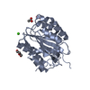 4c29C  4c2aSC C: citing same article ( S: Starting model for refinement |
|---|---|
| Similar structure data |

-Links
 PDBj
PDBj


- Assembly
| Deposited unit | 
| ||||||||
|---|---|---|---|---|---|---|---|---|---|
| 1 | 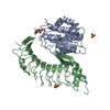
| ||||||||
| 2 | 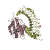
| ||||||||
| 3 | 
| ||||||||
| 4 | 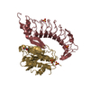
| ||||||||
| Unit cell |
|
-Components
-Protein , 2 types, 8 molecules ACEGBDFH
| #1: Protein | Mass: 24706.678 Da / Num. of mol.: 4 / Fragment: 1264-1468 / Mutation: YES Source method: isolated from a genetically manipulated source Details: HIGH AFFINITY MUTANT OF A1 / Source: (gene. exp.) HOMO SAPIENS (human) / Production host:  #2: Protein | Mass: 32492.104 Da / Num. of mol.: 4 / Mutation: YES Source method: isolated from a genetically manipulated source Details: HIGH AFFINITY MUTANT OF GPIB ALPHA WITH MUTATIONS TO REMOVE N-LINKED GLYCOSYLATION SITES, AND TWO PLATELET TYPE VWD MUTATIONS. Source: (gene. exp.) HOMO SAPIENS (human) / Plasmid: ET-6 / Cell line (production host): HEK-293T / Production host: HOMO SAPIENS (human) / References: UniProt: P07359 |
|---|
-Non-polymers , 4 types, 77 molecules 


| #3: Chemical | ChemComp-SO4 /  Mass: 96.063 Da / Num. of mol.: 10 / Source method: obtained synthetically / Formula: SO4 Mass: 96.063 Da / Num. of mol.: 10 / Source method: obtained synthetically / Formula: SO4#4: Chemical | ChemComp-MES / |  Mass: 195.237 Da / Num. of mol.: 1 / Source method: obtained synthetically / Formula: C6H13NO4S / Comment: pH buffer*YM Mass: 195.237 Da / Num. of mol.: 1 / Source method: obtained synthetically / Formula: C6H13NO4S / Comment: pH buffer*YM#5: Chemical | ChemComp-PEG / |  Mass: 106.120 Da / Num. of mol.: 1 / Source method: obtained synthetically / Formula: C4H10O3 Mass: 106.120 Da / Num. of mol.: 1 / Source method: obtained synthetically / Formula: C4H10O3#6: Water | ChemComp-HOH / | Mass: 18.015 Da / Num. of mol.: 65 / Source method: isolated from a natural source / Formula: H2O |
|---|
-Details
| Has protein modification | Y |
|---|
-Experimental details
-Experiment
| Experiment | Method: X-RAY DIFFRACTION / Number of used crystals: 1 |
|---|
- Sample preparation
| Crystal | Density Matthews: 2.57 Å3/Da / Density % sol: 52.23 % Description: THE RESOLUTION FOR A1 SS-GPIBALPHA-VWD2 WAS FOUND TO EXTEND TO 2.8 ANGSTROMS USING CROSS-CORRELATION. |
|---|---|
| Crystal grow | pH: 4.6 Details: CRYSTALS OF A1/SS-GPIB/VWD2 COMPLEX APPEARED IN DROPS WITH 20% PEG 4000, 0.16 M AMMONIUM SULFATE, 0.08M SODIUM ACETATE, PH 4.6, AND 20% GLYCEROL. THESE CRYSTALS WERE CRUSHED AND USED FOR ...Details: CRYSTALS OF A1/SS-GPIB/VWD2 COMPLEX APPEARED IN DROPS WITH 20% PEG 4000, 0.16 M AMMONIUM SULFATE, 0.08M SODIUM ACETATE, PH 4.6, AND 20% GLYCEROL. THESE CRYSTALS WERE CRUSHED AND USED FOR SEEDING CRYSTAL GROWTH IN 8 MG/ML COMPLEX, 15% PEG 4000, 0.16 M AMMONIUM SULFATE, 0.08 M SODIUM ACETATE, PH 4.6, AND 20% GLYCEROL. SINCE THESE CRYSTALS WERE FORMED IN BUFFER CONTAINING 20% GLYCEROL, NO ADDITIONAL CRYOPROTECTION WAS USED. |
-Data collection
| Diffraction | Mean temperature: 93 K |
|---|---|
| Diffraction source | Source: SYNCHROTRON / Site: APS  / Beamline: 22-ID / Wavelength: 1 / Beamline: 22-ID / Wavelength: 1 |
| Detector | Type: MARRESEARCH MX-300 / Detector: CCD / Date: Apr 5, 2013 |
| Radiation | Protocol: SINGLE WAVELENGTH / Monochromatic (M) / Laue (L): M / Scattering type: x-ray |
| Radiation wavelength | Wavelength: 1 Å / Relative weight: 1 |
| Reflection twin | Operator: h,-k,-l / Fraction: 0.48 |
| Reflection | Resolution: 2.8→48.3 Å / Num. obs: 56526 / % possible obs: 96.6 % / Observed criterion σ(I): -3 / Redundancy: 2.5 % / Rmerge(I) obs: 0.59 / Net I/σ(I): 2.56 |
| Reflection shell | Resolution: 2.8→2.87 Å / Mean I/σ(I) obs: 0.3 / % possible all: 80.9 |
- Processing
| Software |
| |||||||||||||||||||||||||||||||||||||||||||||||||||||||||||||||||||||||||||||||||||||||||||||||||||||||||||||||||||||||||||||||||||||||||||||||||||||||||||||||||||||||||||||||||||||||||||||||||||||||||||||||||||||||||||||||||
|---|---|---|---|---|---|---|---|---|---|---|---|---|---|---|---|---|---|---|---|---|---|---|---|---|---|---|---|---|---|---|---|---|---|---|---|---|---|---|---|---|---|---|---|---|---|---|---|---|---|---|---|---|---|---|---|---|---|---|---|---|---|---|---|---|---|---|---|---|---|---|---|---|---|---|---|---|---|---|---|---|---|---|---|---|---|---|---|---|---|---|---|---|---|---|---|---|---|---|---|---|---|---|---|---|---|---|---|---|---|---|---|---|---|---|---|---|---|---|---|---|---|---|---|---|---|---|---|---|---|---|---|---|---|---|---|---|---|---|---|---|---|---|---|---|---|---|---|---|---|---|---|---|---|---|---|---|---|---|---|---|---|---|---|---|---|---|---|---|---|---|---|---|---|---|---|---|---|---|---|---|---|---|---|---|---|---|---|---|---|---|---|---|---|---|---|---|---|---|---|---|---|---|---|---|---|---|---|---|---|---|---|---|---|---|---|---|---|---|---|---|---|---|---|---|---|---|
| Refinement | Method to determine structure: MOLECULAR REPLACEMENT Starting model: PDB ENTRY 4C2A Resolution: 2.8→48.265 Å / SU ML: 0 / σ(F): 2 / Phase error: 27.94 / Stereochemistry target values: TWIN_LSQ_F
| |||||||||||||||||||||||||||||||||||||||||||||||||||||||||||||||||||||||||||||||||||||||||||||||||||||||||||||||||||||||||||||||||||||||||||||||||||||||||||||||||||||||||||||||||||||||||||||||||||||||||||||||||||||||||||||||||
| Solvent computation | Shrinkage radii: 0.9 Å / VDW probe radii: 1.11 Å / Solvent model: FLAT BULK SOLVENT MODEL | |||||||||||||||||||||||||||||||||||||||||||||||||||||||||||||||||||||||||||||||||||||||||||||||||||||||||||||||||||||||||||||||||||||||||||||||||||||||||||||||||||||||||||||||||||||||||||||||||||||||||||||||||||||||||||||||||
| Refinement step | Cycle: LAST / Resolution: 2.8→48.265 Å
| |||||||||||||||||||||||||||||||||||||||||||||||||||||||||||||||||||||||||||||||||||||||||||||||||||||||||||||||||||||||||||||||||||||||||||||||||||||||||||||||||||||||||||||||||||||||||||||||||||||||||||||||||||||||||||||||||
| Refine LS restraints |
| |||||||||||||||||||||||||||||||||||||||||||||||||||||||||||||||||||||||||||||||||||||||||||||||||||||||||||||||||||||||||||||||||||||||||||||||||||||||||||||||||||||||||||||||||||||||||||||||||||||||||||||||||||||||||||||||||
| LS refinement shell |
| |||||||||||||||||||||||||||||||||||||||||||||||||||||||||||||||||||||||||||||||||||||||||||||||||||||||||||||||||||||||||||||||||||||||||||||||||||||||||||||||||||||||||||||||||||||||||||||||||||||||||||||||||||||||||||||||||
| Refinement TLS params. | Method: refined / Refine-ID: X-RAY DIFFRACTION
| |||||||||||||||||||||||||||||||||||||||||||||||||||||||||||||||||||||||||||||||||||||||||||||||||||||||||||||||||||||||||||||||||||||||||||||||||||||||||||||||||||||||||||||||||||||||||||||||||||||||||||||||||||||||||||||||||
| Refinement TLS group |
|
