 Movie
Movie Controller

[English] 日本語
 Yorodumi
- PDB-4ki3: 1.70 Angstrom resolution crystal structure of outer-membrane lipo... -
+ Open data

- Basic information
| Entry | Database: PDB / ID: 4ki3 | ||||||
|---|---|---|---|---|---|---|---|
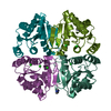
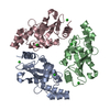
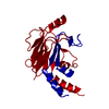
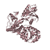
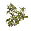
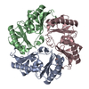
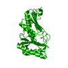
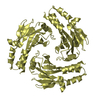
| Title | 1.70 Angstrom resolution crystal structure of outer-membrane lipoprotein carrier protein (lolA) from Yersinia pestis CO92 | ||||||
 Components Components | Outer-membrane lipoprotein carrier protein | ||||||
 Keywords Keywords | CHAPERONE / IDP02066 / Center for Structural Genomics of Infectious Diseases / CSGID / NIAID / National Institute of Allergy and Infectious Diseases | ||||||
| Function / homology | Lipoprotein localisation LolA/LolB/LppX / outer membrane lipoprotein receptor (LolB), chain A / Clam / Mainly Beta / ACETATE ION / DI(HYDROXYETHYL)ETHER / :  Function and homology information Function and homology information | ||||||
| Biological species |  Yersinia pestis biovar Medievalis (bacteria) Yersinia pestis biovar Medievalis (bacteria) | ||||||
| Method |  X-RAY DIFFRACTION / SYNCHROTRON / MOLECULAR REPLACEMENT / Resolution: 1.7 Å X-RAY DIFFRACTION / SYNCHROTRON / MOLECULAR REPLACEMENT / Resolution: 1.7 Å | ||||||
 Authors Authors | Halavaty, A.S. / Wawrzak, Z. / Kudritska, M. / Savchenko, A. / Anderson, W.F. / Center for Structural Genomics of Infectious Diseases (CSGID) | ||||||
 Citation Citation | Journal: To be Published Title: 1.70 Angstrom resolution crystal structure of outer-membrane lipoprotein carrier protein (lolA) from Yersinia pestis CO92 Authors: Halavaty, A.S. / Wawrzak, Z. / Kudritska, M. / Savchenko, A. / Anderson, W.F. / Center for Structural Genomics of Infectious Diseases (CSGID) | ||||||
| History |
|
- Structure visualization
| Structure viewer | Molecule: MolmilJmol/JSmol |
|---|
- Downloads & links
-Download
| PDBx/mmCIF format | 4ki3.cif.gz | 465.6 KB | Display | PDBx/mmCIF format |
|---|---|---|---|---|
| PDB format | pdb4ki3.ent.gz | 383.1 KB | Display | PDB format |
| PDBx/mmJSON format | 4ki3.json.gz | Tree view | PDBx/mmJSON format | |
| Others |  Other downloads Other downloads |
-Validation report
| Arichive directory | https://data.pdbj.org/pub/pdb/validation_reports/ki/4ki3ftp://data.pdbj.org/pub/pdb/validation_reports/ki/4ki3 | HTTPS FTP |
|---|
-Related structure data
| Related structure data |  3ksnS S: Starting model for refinement |
|---|---|
| Similar structure data | |
| Other databases |
-Links
 PDBj
PDBj
- Assembly
| Deposited unit | 
| ||||||||
|---|---|---|---|---|---|---|---|---|---|
| 1 | 
| ||||||||
| 2 | 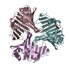
| ||||||||
| 3 | 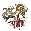
| ||||||||
| 4 | 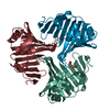
| ||||||||
| Unit cell |
|
-Components
| #1: Protein | Mass: 20383.354 Da / Num. of mol.: 12 Source method: isolated from a genetically manipulated source Source: (gene. exp.) Yersinia pestis biovar Medievalis (bacteria)Strain: Harbin 35 / Gene: lolA, YPC_2798 / Plasmid: pMCSG53 / Production host: #2: Chemical | ChemComp-ACT /   Mass: 59.044 Da / Num. of mol.: 7 / Source method: obtained synthetically / Formula: C2H3O2 Mass: 59.044 Da / Num. of mol.: 7 / Source method: obtained synthetically / Formula: C2H3O2#3: Chemical |   Mass: 106.120 Da / Num. of mol.: 3 / Source method: obtained synthetically / Formula: C4H10O3 Mass: 106.120 Da / Num. of mol.: 3 / Source method: obtained synthetically / Formula: C4H10O3#4: Chemical |   Mass: 92.094 Da / Num. of mol.: 2 / Source method: obtained synthetically / Formula: C3H8O3 Mass: 92.094 Da / Num. of mol.: 2 / Source method: obtained synthetically / Formula: C3H8O3#5: Water | ChemComp-HOH / |  Mass: 18.015 Da / Num. of mol.: 1497 / Source method: isolated from a natural source / Formula: H2O Mass: 18.015 Da / Num. of mol.: 1497 / Source method: isolated from a natural source / Formula: H2O |
|---|
-Experimental details
-Experiment
| Experiment | Method: X-RAY DIFFRACTION / Number of used crystals: 1 |
|---|
- Sample preparation
| Crystal | Density Matthews: 2.09 Å3/Da / Density % sol: 41.28 % |
|---|---|
| Crystal grow | Temperature: 295 K / Method: vapor diffusion, hanging drop / pH: 7 Details: PEG 3350 20% w/v NH4 dihydrogen Phosphate 0.2M, protein at 34 mg/mL, cryo- 20% glycerol+paratone, pH 7, VAPOR DIFFUSION, HANGING DROP, temperature 295K |
-Data collection
| Diffraction | Mean temperature: 100 K | |||||||||||||||
|---|---|---|---|---|---|---|---|---|---|---|---|---|---|---|---|---|
| Diffraction source | Source: SYNCHROTRON / Site: APS  / Beamline: 21-ID-G / Wavelength: 0.97856 Å / Beamline: 21-ID-G / Wavelength: 0.97856 Å | |||||||||||||||
| Detector | Type: MARMOSAIC 300 mm CCD / Detector: CCD / Date: Feb 9, 2013 / Details: Be-Lenses | |||||||||||||||
| Radiation | Monochromator: Diamond / Protocol: SINGLE WAVELENGTH / Monochromatic (M) / Laue (L): M / Scattering type: x-ray | |||||||||||||||
| Radiation wavelength | Wavelength: 0.97856 Å / Relative weight: 1 | |||||||||||||||
| Reflection twin |
| |||||||||||||||
| Reflection | Resolution: 1.7→30 Å / Num. all: 207123 / Num. obs: 207123 / % possible obs: 94.8 % / Observed criterion σ(I): -3 / Redundancy: 4.1 % / Biso Wilson estimate: 23.3 Å2 / Rmerge(I) obs: 0.091 / Net I/σ(I): 14.22 | |||||||||||||||
| Reflection shell | Resolution: 1.7→1.73 Å / Redundancy: 4.1 % / Rmerge(I) obs: 0.598 / Mean I/σ(I) obs: 2.7 / Num. unique all: 10516 / % possible all: 95.4 |
- Processing
| Software |
| |||||||||||||||||||||||||||||||||||||||||||||||||||||||||||||||||||||||||||||||||||||
|---|---|---|---|---|---|---|---|---|---|---|---|---|---|---|---|---|---|---|---|---|---|---|---|---|---|---|---|---|---|---|---|---|---|---|---|---|---|---|---|---|---|---|---|---|---|---|---|---|---|---|---|---|---|---|---|---|---|---|---|---|---|---|---|---|---|---|---|---|---|---|---|---|---|---|---|---|---|---|---|---|---|---|---|---|---|---|
| Refinement | Method to determine structure: MOLECULAR REPLACEMENT Starting model: PDB entry 3KSN Resolution: 1.7→29.09 Å / Cor.coef. Fo:Fc: 0.949 / Cor.coef. Fo:Fc free: 0.933 / SU B: 2.607 / SU ML: 0.082 / Isotropic thermal model: Isotropic / Cross valid method: THROUGHOUT / ESU R: 0.03 / ESU R Free: 0.028 / Stereochemistry target values: MAXIMUM LIKELIHOOD / Details: HYDROGENS HAVE BEEN ADDED IN THE RIDING POSITIONS
| |||||||||||||||||||||||||||||||||||||||||||||||||||||||||||||||||||||||||||||||||||||
| Solvent computation | Ion probe radii: 0.8 Å / Shrinkage radii: 0.8 Å / VDW probe radii: 1.2 Å / Solvent model: BABINET MODEL WITH MASK | |||||||||||||||||||||||||||||||||||||||||||||||||||||||||||||||||||||||||||||||||||||
| Displacement parameters | Biso mean: 22.516 Å2
| |||||||||||||||||||||||||||||||||||||||||||||||||||||||||||||||||||||||||||||||||||||
| Refinement step | Cycle: LAST / Resolution: 1.7→29.09 Å
| |||||||||||||||||||||||||||||||||||||||||||||||||||||||||||||||||||||||||||||||||||||
| Refine LS restraints |
| |||||||||||||||||||||||||||||||||||||||||||||||||||||||||||||||||||||||||||||||||||||
| LS refinement shell | Resolution: 1.7→1.738 Å / Total num. of bins used: 20
|
