 Movie
Movie Controller


+ Open data

- Basic information
| Entry | Database: PDB / ID: 3ksn | ||||||
|---|---|---|---|---|---|---|---|
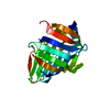
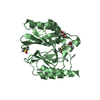
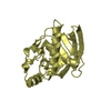
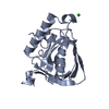
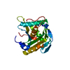
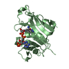
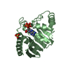
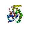
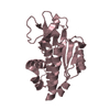
| Title | Crystal structure of the lipoprotein localization factor, LolA | ||||||
 Components Components | Outer-membrane lipoprotein carrier protein | ||||||
 Keywords Keywords | PROTEIN TRANSPORT / lipoprotein carrier / Chaperone / Transport | ||||||
| Function / homology |  Function and homology information Function and homology informationlipoprotein localization to outer membrane / lipoprotein transport / outer membrane-bounded periplasmic space Similarity search - Function | ||||||
| Biological species |  | ||||||
| Method |  X-RAY DIFFRACTION / SYNCHROTRON / MOLECULAR REPLACEMENT / Resolution: 1.65 Å X-RAY DIFFRACTION / SYNCHROTRON / MOLECULAR REPLACEMENT / Resolution: 1.65 Å | ||||||
 Authors Authors | Ahmadpour, F. / Gloyd, M. / Guarne, A. / Stewart, G. / Pathania, R. / Brown, E.D. | ||||||
 Citation Citation | Journal: To be Published Title: Crystal structure of the lipoprotein localization factor, LolA Authors: Ahmadpour, F. / Gloyd, M. / Guarne, A. / Stewart, G. / Pathania, R. / Brown, E.D. | ||||||
| History |
|
- Structure visualization
| Structure viewer | Molecule: MolmilJmol/JSmol |
|---|
- Downloads & links
-Download
| PDBx/mmCIF format | 3ksn.cif.gz | 85.7 KB | Display | PDBx/mmCIF format |
|---|---|---|---|---|
| PDB format | pdb3ksn.ent.gz | 64.5 KB | Display | PDB format |
| PDBx/mmJSON format | 3ksn.json.gz | Tree view | PDBx/mmJSON format | |
| Others |  Other downloads Other downloads |
-Validation report
| Arichive directory | https://data.pdbj.org/pub/pdb/validation_reports/ks/3ksnftp://data.pdbj.org/pub/pdb/validation_reports/ks/3ksn | HTTPS FTP |
|---|
-Related structure data
| Related structure data |  1au8S S: Starting model for refinement |
|---|---|
| Similar structure data |


-Links
 PDBj
PDBj
- Assembly
| Deposited unit | 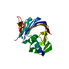
| ||||||||||||
|---|---|---|---|---|---|---|---|---|---|---|---|---|---|
| 1 |
| ||||||||||||
| Unit cell |
| ||||||||||||
| Components on special symmetry positions |
|
-Components
| #1: Protein | Mass: 20400.348 Da / Num. of mol.: 1 / Fragment: Residues 22-203 Source method: isolated from a genetically manipulated source Source: (gene. exp.) |
|---|---|
| #2: Water | ChemComp-HOH /  Mass: 18.015 Da / Num. of mol.: 79 / Source method: isolated from a natural source / Formula: H2O Mass: 18.015 Da / Num. of mol.: 79 / Source method: isolated from a natural source / Formula: H2O |
-Experimental details
-Experiment
| Experiment | Method: X-RAY DIFFRACTION / Number of used crystals: 1 |
|---|
- Sample preparation
| Crystal | Density Matthews: 2.04 Å3/Da / Density % sol: 39.78 % |
|---|---|
| Crystal grow | Temperature: 277 K / Method: vapor diffusion, hanging drop / pH: 4.4 Details: 25% PEG 1000, 20% PEG 8000, 0.1 M sodium acetate, dehydrated over 1M ammonium sulfate, pH 4.4, VAPOR DIFFUSION, HANGING DROP, temperature 277K |
-Data collection
| Diffraction | Mean temperature: 100 K |
|---|---|
| Diffraction source | Source: SYNCHROTRON / Site: NSLS  / Beamline: X29A / Wavelength: 1.0809 Å / Beamline: X29A / Wavelength: 1.0809 Å |
| Detector | Type: ADSC QUANTUM 315r / Detector: CCD / Date: Apr 24, 2008 / Details: mirrors |
| Radiation | Monochromator: Si(111) / Protocol: SINGLE WAVELENGTH / Monochromatic (M) / Laue (L): M / Scattering type: x-ray |
| Radiation wavelength | Wavelength: 1.0809 Å / Relative weight: 1 |
| Reflection | Resolution: 1.65→50 Å / Num. obs: 20611 / % possible obs: 99.6 % / Redundancy: 9.8 % / Biso Wilson estimate: 23.11 Å2 / Rmerge(I) obs: 0.047 / Rsym value: 0.035 / Net I/σ(I): 39.5 |
| Reflection shell | Resolution: 1.65→1.71 Å / Redundancy: 9.8 % / Rmerge(I) obs: 0.534 / Mean I/σ(I) obs: 4.47 / Num. unique all: 2035 / Rsym value: 0.491 / % possible all: 99.7 |
- Processing
| Software |
| |||||||||||||||||||||||||||||||||||||||||||||||||||||||||||||||
|---|---|---|---|---|---|---|---|---|---|---|---|---|---|---|---|---|---|---|---|---|---|---|---|---|---|---|---|---|---|---|---|---|---|---|---|---|---|---|---|---|---|---|---|---|---|---|---|---|---|---|---|---|---|---|---|---|---|---|---|---|---|---|---|---|
| Refinement | Method to determine structure: MOLECULAR REPLACEMENT Starting model: 1AU8 Resolution: 1.65→31.577 Å / SU ML: 0.24 / Cross valid method: THROUGHOUT / σ(F): 1.89 / Stereochemistry target values: ML
| |||||||||||||||||||||||||||||||||||||||||||||||||||||||||||||||
| Solvent computation | Shrinkage radii: 0.9 Å / VDW probe radii: 1.11 Å / Solvent model: FLAT BULK SOLVENT MODEL / Bsol: 60.497 Å2 / ksol: 0.4 e/Å3 | |||||||||||||||||||||||||||||||||||||||||||||||||||||||||||||||
| Displacement parameters |
| |||||||||||||||||||||||||||||||||||||||||||||||||||||||||||||||
| Refinement step | Cycle: LAST / Resolution: 1.65→31.577 Å
| |||||||||||||||||||||||||||||||||||||||||||||||||||||||||||||||
| Refine LS restraints |
| |||||||||||||||||||||||||||||||||||||||||||||||||||||||||||||||
| LS refinement shell | Refine-ID: X-RAY DIFFRACTION
|
