 Movie
Movie Controller

[English] 日本語
 Yorodumi
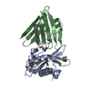
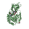
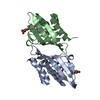
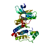
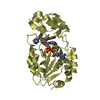
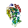
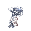
- PDB-4bws: Crystal structure of the heterotrimer of PQBP1, U5-15kD and U5-52kD. -
+ Open data

- Basic information
| Entry | Database: PDB / ID: 4bws | ||||||
|---|---|---|---|---|---|---|---|
| Title | Crystal structure of the heterotrimer of PQBP1, U5-15kD and U5-52kD. | ||||||
 Components Components |
| ||||||
 Keywords Keywords | TRANSCRIPTION / NEURODEGENERATIVE DISORDERS | ||||||
| Function / homology |  Function and homology information Function and homology informationneuronal ribonucleoprotein granule / alternative mRNA splicing, via spliceosome / RNA splicing, via transesterification reactions / U2-type precatalytic spliceosome / cellular response to exogenous dsRNA / regulation of dendrite morphogenesis / regulation of RNA splicing / mRNA Splicing - Minor Pathway / spliceosomal complex assembly / spliceosomal tri-snRNP complex assembly ...neuronal ribonucleoprotein granule / alternative mRNA splicing, via spliceosome / RNA splicing, via transesterification reactions / U2-type precatalytic spliceosome / cellular response to exogenous dsRNA / regulation of dendrite morphogenesis / regulation of RNA splicing / mRNA Splicing - Minor Pathway / spliceosomal complex assembly / spliceosomal tri-snRNP complex assembly / U5 snRNP / ribonucleoprotein complex binding / U4/U6 x U5 tri-snRNP complex / positive regulation of defense response to virus by host / positive regulation of type I interferon production / mRNA Splicing - Major Pathway / activation of innate immune response / spliceosomal complex / mRNA splicing, via spliceosome / fibrillar center / neuron projection development / cytoplasmic stress granule / microtubule cytoskeleton / double-stranded DNA binding / defense response to virus / transcription coactivator activity / nuclear speck / cilium / nuclear body / ciliary basal body / innate immune response / cell division / regulation of DNA-templated transcription / DNA binding / nucleoplasm / nucleus / cytoplasm / cytosol Similarity search - Function | ||||||
| Biological species |  HOMO SAPIENS (human) HOMO SAPIENS (human) | ||||||
| Method |  X-RAY DIFFRACTION / SYNCHROTRON / MOLECULAR REPLACEMENT / Resolution: 2.5 Å X-RAY DIFFRACTION / SYNCHROTRON / MOLECULAR REPLACEMENT / Resolution: 2.5 Å | ||||||
 Authors Authors | Mizuguchi, M. / Obita, T. / Serita, T. / Kojima, R. / Morimoto, T. / Nabeshima, Y. / Okazawa, H. | ||||||
 Citation Citation | Journal: Nat.Commun. / Year: 2014 Title: Mutations in the Pqbp1 Gene Prevent its Interaction with the Spliceosomal Protein U5-15Kd. Authors: Mizuguchi, M. / Obita, T. / Serita, T. / Kojima, R. / Nabeshima, Y. / Okazawa, H. | ||||||
| History |
|
- Structure visualization
| Structure viewer | Molecule: MolmilJmol/JSmol |
|---|
- Downloads & links
-Download
| PDBx/mmCIF format | 4bws.cif.gz | 100.8 KB | Display | PDBx/mmCIF format |
|---|---|---|---|---|
| PDB format | pdb4bws.ent.gz | 77.7 KB | Display | PDB format |
| PDBx/mmJSON format | 4bws.json.gz | Tree view | PDBx/mmJSON format | |
| Others |  Other downloads Other downloads |
-Validation report
| Arichive directory | https://data.pdbj.org/pub/pdb/validation_reports/bw/4bwsftp://data.pdbj.org/pub/pdb/validation_reports/bw/4bws | HTTPS FTP |
|---|
-Related structure data
| Related structure data |  4bwqC  4cdoC  1syxS C: citing same article ( S: Starting model for refinement |
|---|---|
| Similar structure data |


-Links
 PDBj
PDBj


- Assembly
| Deposited unit | 
| ||||||||
|---|---|---|---|---|---|---|---|---|---|
| 1 | 
| ||||||||
| 2 | 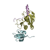
| ||||||||
| Unit cell |
|
-Components
| #1: Protein | Mass: 16743.229 Da / Num. of mol.: 2 / Fragment: RESIDUES 4-137 Source method: isolated from a genetically manipulated source Source: (gene. exp.) HOMO SAPIENS (human) / Production host:  #2: Protein/peptide | Mass: 3876.208 Da / Num. of mol.: 2 / Fragment: RESIDUES 223-265 Source method: isolated from a genetically manipulated source Source: (gene. exp.) HOMO SAPIENS (human) / Production host: #3: Protein | Mass: 8482.363 Da / Num. of mol.: 2 / Fragment: RESIDUES 280-341 Source method: isolated from a genetically manipulated source Source: (gene. exp.) HOMO SAPIENS (human) / Plasmid: POPTH / Production host: #4: Water | ChemComp-HOH / |  Mass: 18.015 Da / Num. of mol.: 16 / Source method: isolated from a natural source / Formula: H2O Mass: 18.015 Da / Num. of mol.: 16 / Source method: isolated from a natural source / Formula: H2O |
|---|
-Experimental details
-Experiment
| Experiment | Method: X-RAY DIFFRACTION / Number of used crystals: 1 |
|---|
- Sample preparation
| Crystal | Density Matthews: 4.2 Å3/Da / Density % sol: 70 % / Description: NONE |
|---|
-Data collection
| Diffraction | Mean temperature: 100 K |
|---|---|
| Diffraction source | Source: SYNCHROTRON / Site: SPring-8  / Beamline: BL41XU / Wavelength: 1 / Beamline: BL41XU / Wavelength: 1 |
| Detector | Type: MARRESEARCH / Detector: CCD / Date: Oct 27, 2012 |
| Radiation | Protocol: SINGLE WAVELENGTH / Monochromatic (M) / Laue (L): M / Scattering type: x-ray |
| Radiation wavelength | Wavelength: 1 Å / Relative weight: 1 |
| Reflection | Resolution: 2.5→47.27 Å / Num. obs: 34203 / % possible obs: 100 % / Observed criterion σ(I): 1 / Redundancy: 3.6 % / Biso Wilson estimate: 25.28 Å2 / Rmerge(I) obs: 0.24 / Net I/σ(I): 4.1 |
| Reflection shell | Resolution: 2.5→2.64 Å / Redundancy: 3.6 % / Rmerge(I) obs: 0.61 / Mean I/σ(I) obs: 1.8 / % possible all: 100 |
- Processing
| Software |
| |||||||||||||||||||||||||||||||||||||||||||||||||||||||||||||||||||||||||||||||||||||||||||
|---|---|---|---|---|---|---|---|---|---|---|---|---|---|---|---|---|---|---|---|---|---|---|---|---|---|---|---|---|---|---|---|---|---|---|---|---|---|---|---|---|---|---|---|---|---|---|---|---|---|---|---|---|---|---|---|---|---|---|---|---|---|---|---|---|---|---|---|---|---|---|---|---|---|---|---|---|---|---|---|---|---|---|---|---|---|---|---|---|---|---|---|---|
| Refinement | Method to determine structure: MOLECULAR REPLACEMENT Starting model: PDB ENTRY 1SYX Resolution: 2.5→41.818 Å / SU ML: 0.33 / σ(F): 1.36 / Phase error: 30.17 / Stereochemistry target values: ML
| |||||||||||||||||||||||||||||||||||||||||||||||||||||||||||||||||||||||||||||||||||||||||||
| Solvent computation | Shrinkage radii: 0.9 Å / VDW probe radii: 1.11 Å / Solvent model: FLAT BULK SOLVENT MODEL | |||||||||||||||||||||||||||||||||||||||||||||||||||||||||||||||||||||||||||||||||||||||||||
| Refinement step | Cycle: LAST / Resolution: 2.5→41.818 Å
| |||||||||||||||||||||||||||||||||||||||||||||||||||||||||||||||||||||||||||||||||||||||||||
| Refine LS restraints |
| |||||||||||||||||||||||||||||||||||||||||||||||||||||||||||||||||||||||||||||||||||||||||||
| LS refinement shell |
|
