 Movie
Movie Controller

[English] 日本語
 Yorodumi
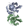
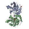
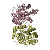
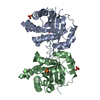
- PDB-4bi6: CRYSTAL STRUCTURE OF A TRIPLE MUTANT (A198V, C202A AND C222N) OF ... -
+ Open data

- Basic information
| Entry | Database: PDB / ID: 4bi6 | ||||||||||||
|---|---|---|---|---|---|---|---|---|---|---|---|---|---|
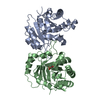
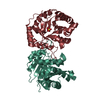
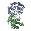
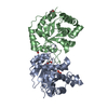
| Title | CRYSTAL STRUCTURE OF A TRIPLE MUTANT (A198V, C202A AND C222N) OF TRIOSEPHOSPHATE ISOMERASE FROM GIARDIA LAMBLIA. COMPLEXED WITH 2- PHOSPHOGLYCOLIC ACID | ||||||||||||
 Components Components | TRIOSEPHOSPHATE ISOMERASE | ||||||||||||
 Keywords Keywords | ISOMERASE | ||||||||||||
| Function / homology |  Function and homology information Function and homology informationmethylglyoxal synthase / methylglyoxal synthase activity / triose-phosphate isomerase / triose-phosphate isomerase activity / glyceraldehyde-3-phosphate biosynthetic process / glycerol catabolic process / glycolytic process / gluconeogenesis / cytosol Similarity search - Function | ||||||||||||
| Biological species |  GIARDIA INTESTINALIS (eukaryote) GIARDIA INTESTINALIS (eukaryote) | ||||||||||||
| Method |  X-RAY DIFFRACTION / SYNCHROTRON / MOLECULAR REPLACEMENT / Resolution: 1.45 Å X-RAY DIFFRACTION / SYNCHROTRON / MOLECULAR REPLACEMENT / Resolution: 1.45 Å | ||||||||||||
 Authors Authors | Torres-Larios, A. / Enriquez-Flores, S. / Reyes-Vivas, H. / Oria-Hernandez, J. / Hernandez-Alcantara, G. | ||||||||||||
 Citation Citation | Journal: Plos One / Year: 2013 Title: Structural and Functional Perturbation of Giardia Lamblia Triosephosphate Isomerase by Modification of a Non-Catalytic, Non-Conserved Region. Authors: Hernandez-Alcantara, G. / Torres-Larios, A. / Enriquez-Flores, S. / Garcia-Torres, I. / Castillo-Villanueva, A. / Mendez, S.T. / De La Mora-De La Mora, I. / Gomez-Manzo, S. / Torres-Arroyo, ...Authors: Hernandez-Alcantara, G. / Torres-Larios, A. / Enriquez-Flores, S. / Garcia-Torres, I. / Castillo-Villanueva, A. / Mendez, S.T. / De La Mora-De La Mora, I. / Gomez-Manzo, S. / Torres-Arroyo, A. / Lopez-Velazquez, G. / Reyes-Vivas, H. / Oria-Hernandez, J. | ||||||||||||
| History |
| ||||||||||||
| Remark 700 | SHEET DETERMINATION METHOD: DSSP THE SHEETS PRESENTED AS "AA" IN EACH CHAIN ON SHEET RECORDS BELOW ... SHEET DETERMINATION METHOD: DSSP THE SHEETS PRESENTED AS "AA" IN EACH CHAIN ON SHEET RECORDS BELOW IS ACTUALLY AN 8-STRANDED BARREL THIS IS REPRESENTED BY A 9-STRANDED SHEET IN WHICH THE FIRST AND LAST STRANDS ARE IDENTICAL. |
- Structure visualization
| Structure viewer | Molecule: MolmilJmol/JSmol |
|---|
- Downloads & links
-Download
| PDBx/mmCIF format | 4bi6.cif.gz | 67.7 KB | Display | PDBx/mmCIF format |
|---|---|---|---|---|
| PDB format | pdb4bi6.ent.gz | 50.1 KB | Display | PDB format |
| PDBx/mmJSON format | 4bi6.json.gz | Tree view | PDBx/mmJSON format | |
| Others |  Other downloads Other downloads |
-Validation report
| Arichive directory | https://data.pdbj.org/pub/pdb/validation_reports/bi/4bi6ftp://data.pdbj.org/pub/pdb/validation_reports/bi/4bi6 | HTTPS FTP |
|---|
-Related structure data
| Related structure data |  4bi5C  4bi7C  2dp3S C: citing same article ( S: Starting model for refinement |
|---|---|
| Similar structure data |


-Links
 PDBj
PDBj
- Assembly
| Deposited unit | 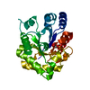
| ||||||||||||||||||
|---|---|---|---|---|---|---|---|---|---|---|---|---|---|---|---|---|---|---|---|
| 1 | 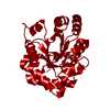
| ||||||||||||||||||
| Unit cell |
| ||||||||||||||||||
| Components on special symmetry positions |
|
-Components
| #1: Protein | Mass: 27947.135 Da / Num. of mol.: 1 / Mutation: YES Source method: isolated from a genetically manipulated source Source: (gene. exp.) GIARDIA INTESTINALIS (eukaryote) / Production host:  |
|---|---|
| #2: Chemical | ChemComp-PGA / 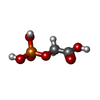  Mass: 156.031 Da / Num. of mol.: 1 Mass: 156.031 Da / Num. of mol.: 1Source method: isolated from a genetically manipulated source Formula: C2H5O6P |
| #3: Water | ChemComp-HOH /  Mass: 18.015 Da / Num. of mol.: 220 / Source method: isolated from a natural source / Formula: H2O Mass: 18.015 Da / Num. of mol.: 220 / Source method: isolated from a natural source / Formula: H2O |
-Experimental details
-Experiment
| Experiment | Method: X-RAY DIFFRACTION / Number of used crystals: 1 |
|---|
- Sample preparation
| Crystal | Density Matthews: 2.98 Å3/Da / Density % sol: 58.71 % / Description: NONE |
|---|---|
| Crystal grow | pH: 5.6 Details: PROTEIN WAS CRYSTALLYZED FROM 0.5 M SODIUM CHLORIDE, 0.1 M SODIUM CITRATE TRIBASIC DIHYDRATE PH 5.6, 2 % V/V ETHYLENE IMINE POLYMER. |
-Data collection
| Diffraction | Mean temperature: 90 K |
|---|---|
| Diffraction source | Source: SYNCHROTRON / Site: APS  / Beamline: 21-ID-F / Wavelength: 0.97857 / Beamline: 21-ID-F / Wavelength: 0.97857 |
| Detector | Type: MARRESEARCH / Detector: CCD / Date: Nov 5, 2009 / Details: MIRRORS |
| Radiation | Monochromator: DIAMOND / Protocol: SINGLE WAVELENGTH / Monochromatic (M) / Laue (L): M / Scattering type: x-ray |
| Radiation wavelength | Wavelength: 0.97857 Å / Relative weight: 1 |
| Reflection | Resolution: 1.45→37.55 Å / Num. obs: 58919 / % possible obs: 99.9 % / Observed criterion σ(I): 0 / Redundancy: 6.8 % / Rmerge(I) obs: 0.09 / Net I/σ(I): 3.7 |
| Reflection shell | Resolution: 1.45→1.53 Å / Redundancy: 6.8 % / Rmerge(I) obs: 0.35 / Mean I/σ(I) obs: 2.1 / % possible all: 99.9 |
- Processing
| Software |
| ||||||||||||||||||||||||||||||||||||||||||||||||||||||||||||||||||||||||||||||||||||||||||||||||||||||||||||||||||||||||||||||||||||||||||||||||||||||||||||||||||||||||||||||||||||||
|---|---|---|---|---|---|---|---|---|---|---|---|---|---|---|---|---|---|---|---|---|---|---|---|---|---|---|---|---|---|---|---|---|---|---|---|---|---|---|---|---|---|---|---|---|---|---|---|---|---|---|---|---|---|---|---|---|---|---|---|---|---|---|---|---|---|---|---|---|---|---|---|---|---|---|---|---|---|---|---|---|---|---|---|---|---|---|---|---|---|---|---|---|---|---|---|---|---|---|---|---|---|---|---|---|---|---|---|---|---|---|---|---|---|---|---|---|---|---|---|---|---|---|---|---|---|---|---|---|---|---|---|---|---|---|---|---|---|---|---|---|---|---|---|---|---|---|---|---|---|---|---|---|---|---|---|---|---|---|---|---|---|---|---|---|---|---|---|---|---|---|---|---|---|---|---|---|---|---|---|---|---|---|---|
| Refinement | Method to determine structure: MOLECULAR REPLACEMENT Starting model: PDB ENTRY 2DP3 Resolution: 1.45→37.55 Å / Cor.coef. Fo:Fc: 0.959 / Cor.coef. Fo:Fc free: 0.955 / Cross valid method: THROUGHOUT / ESU R: 0.061 / ESU R Free: 0.061 / Stereochemistry target values: MAXIMUM LIKELIHOOD Details: HYDROGENS HAVE BEEN ADDED IN THE RIDING POSITIONS. U VALUES REFINED INDIVIDUALLY
| ||||||||||||||||||||||||||||||||||||||||||||||||||||||||||||||||||||||||||||||||||||||||||||||||||||||||||||||||||||||||||||||||||||||||||||||||||||||||||||||||||||||||||||||||||||||
| Solvent computation | Ion probe radii: 0.8 Å / Shrinkage radii: 0.8 Å / VDW probe radii: 1.4 Å / Solvent model: MASK | ||||||||||||||||||||||||||||||||||||||||||||||||||||||||||||||||||||||||||||||||||||||||||||||||||||||||||||||||||||||||||||||||||||||||||||||||||||||||||||||||||||||||||||||||||||||
| Displacement parameters | Biso mean: 18.761 Å2
| ||||||||||||||||||||||||||||||||||||||||||||||||||||||||||||||||||||||||||||||||||||||||||||||||||||||||||||||||||||||||||||||||||||||||||||||||||||||||||||||||||||||||||||||||||||||
| Refinement step | Cycle: LAST / Resolution: 1.45→37.55 Å
| ||||||||||||||||||||||||||||||||||||||||||||||||||||||||||||||||||||||||||||||||||||||||||||||||||||||||||||||||||||||||||||||||||||||||||||||||||||||||||||||||||||||||||||||||||||||
| Refine LS restraints |
|