 Movie
Movie Controller

[English] 日本語
 Yorodumi
- PDB-3vw3: Antibody 64M-5 Fab in complex with a double-stranded DNA (6-4) ph... -
+ Open data

- Basic information
| Entry | Database: PDB / ID: 3vw3 | ||||||
|---|---|---|---|---|---|---|---|
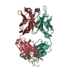
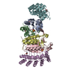
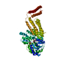
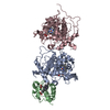
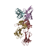
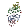
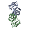
| Title | Antibody 64M-5 Fab in complex with a double-stranded DNA (6-4) photoproduct | ||||||
 Components Components |
| ||||||
 Keywords Keywords | IMMUNE SYSTEM/DNA / Protein-DNA complex / DNA (6-4) photoproduct / Immunoglobulin / IMMUNE SYSTEM-DNA complex | ||||||
| Function / homology | Immunoglobulins / Immunoglobulin-like / Sandwich / Mainly Beta / COBALT HEXAMMINE(III) / DNA / DNA (> 10) Function and homology information Function and homology information | ||||||
| Biological species |  | ||||||
| Method |  X-RAY DIFFRACTION / SYNCHROTRON / MOLECULAR REPLACEMENT / Resolution: 2.5 Å X-RAY DIFFRACTION / SYNCHROTRON / MOLECULAR REPLACEMENT / Resolution: 2.5 Å | ||||||
 Authors Authors | Yokoyama, H. / Mizutani, R. / Satow, Y. | ||||||
 Citation Citation | Journal: Acta Crystallogr.,Sect.D / Year: 2013 Title: Structure of a double-stranded DNA (6-4) photoproduct in complex with the 64M-5 antibody Fab Authors: Yokoyama, H. / Mizutani, R. / Satow, Y. #1: Journal: Acta Crystallogr.,Sect.D / Year: 2012Title: Structure of the DNA (6-4) photoproduct dTT(6-4)TT in complex with the 64M-2 antibody Fab fragment implies increased antibody-binding affinity by the flanking nucleotides Authors: Yokoyama, H. / Mizutani, R. / Satow, Y. / Sato, K. / Komatsu, Y. / Ohtsuka, E. / Nikaido, O. #2: Journal: J.Mol.Biol. / Year: 2000Title: Crystal structure of the 64M-2 antibody Fab fragment in complex with a DNA dT(6-4)T photoproduct formed by ultraviolet radiation Authors: Yokoyama, H. / Mizutani, R. / Satow, Y. / Komatsu, Y. / Ohtsuka, E. / Nikaido, O. | ||||||
| History |
|
- Structure visualization
| Structure viewer | Molecule: MolmilJmol/JSmol |
|---|
- Downloads & links
-Download
| PDBx/mmCIF format | 3vw3.cif.gz | 123.4 KB | Display | PDBx/mmCIF format |
|---|---|---|---|---|
| PDB format | pdb3vw3.ent.gz | 91.2 KB | Display | PDB format |
| PDBx/mmJSON format | 3vw3.json.gz | Tree view | PDBx/mmJSON format | |
| Others |  Other downloads Other downloads |
-Validation report
| Arichive directory | https://data.pdbj.org/pub/pdb/validation_reports/vw/3vw3ftp://data.pdbj.org/pub/pdb/validation_reports/vw/3vw3 | HTTPS FTP |
|---|
-Related structure data
| Related structure data | 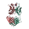 1kegS S: Starting model for refinement |
|---|---|
| Similar structure data |


-Links
 PDBj
PDBj


- Assembly
| Deposited unit | 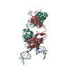
| ||||||||
|---|---|---|---|---|---|---|---|---|---|
| 1 |
| ||||||||
| Unit cell |
|
-Components
-DNA chain , 2 types, 2 molecules AB
| #3: DNA chain | Mass: 5638.668 Da / Num. of mol.: 1 / Source method: obtained synthetically / Details: DNA with (6-4) photoproduct |
|---|---|
| #4: DNA chain | Mass: 5396.507 Da / Num. of mol.: 1 / Source method: obtained synthetically / Details: synthetic DNA |
-Antibody , 2 types, 2 molecules LH
| #1: Antibody | Mass: 23993.572 Da / Num. of mol.: 1 / Source method: isolated from a natural source / Details: hybridoma / Source: (natural) |
|---|---|
| #2: Antibody | Mass: 23713.553 Da / Num. of mol.: 1 / Source method: isolated from a natural source / Details: hybridoma / Source: (natural) |
-Non-polymers , 2 types, 145 molecules 

| #5: Chemical | ChemComp-NCO /  Mass: 161.116 Da / Num. of mol.: 1 / Source method: obtained synthetically / Formula: CoH18N6 Mass: 161.116 Da / Num. of mol.: 1 / Source method: obtained synthetically / Formula: CoH18N6 |
|---|---|
| #6: Water | ChemComp-HOH / Mass: 18.015 Da / Num. of mol.: 144 / Source method: isolated from a natural source / Formula: H2O |
-Details
| Has protein modification | Y |
|---|
-Experimental details
-Experiment
| Experiment | Method: X-RAY DIFFRACTION / Number of used crystals: 1 |
|---|
- Sample preparation
| Crystal | Density Matthews: 2.43 Å3/Da / Density % sol: 49.34 % |
|---|---|
| Crystal grow | Temperature: 277 K / Method: vapor diffusion, hanging drop / pH: 8 Details: 12% PEG 3350, 0.1M magnesium acetate, 10mM Cobalt hexamine chloride, 0.1M Tris-HCl, pH 8.0, VAPOR DIFFUSION, HANGING DROP, temperature 277K |
-Data collection
| Diffraction | Mean temperature: 100 K |
|---|---|
| Diffraction source | Source: SYNCHROTRON / Site: SPring-8  / Beamline: BL41XU / Wavelength: 0.8 Å / Beamline: BL41XU / Wavelength: 0.8 Å |
| Detector | Type: MAR CCD 165 mm / Detector: CCD / Date: Mar 1, 2002 / Details: mirrors |
| Radiation | Monochromator: Si(111) / Protocol: SINGLE WAVELENGTH / Monochromatic (M) / Laue (L): M / Scattering type: x-ray |
| Radiation wavelength | Wavelength: 0.8 Å / Relative weight: 1 |
| Reflection | Resolution: 2.5→30 Å / Num. obs: 20541 / % possible obs: 97.1 % / Redundancy: 10.5 % / Biso Wilson estimate: 49.1 Å2 / Rmerge(I) obs: 0.054 / Net I/σ(I): 60.7 |
| Reflection shell | Resolution: 2.5→2.59 Å / Rmerge(I) obs: 0.24 / Mean I/σ(I) obs: 16 / % possible all: 97.2 |
- Processing
| Software |
| ||||||||||||||||||||||||||||||||||||||||||||||||||||||||||||||||||||||||||||||||
|---|---|---|---|---|---|---|---|---|---|---|---|---|---|---|---|---|---|---|---|---|---|---|---|---|---|---|---|---|---|---|---|---|---|---|---|---|---|---|---|---|---|---|---|---|---|---|---|---|---|---|---|---|---|---|---|---|---|---|---|---|---|---|---|---|---|---|---|---|---|---|---|---|---|---|---|---|---|---|---|---|---|
| Refinement | Method to determine structure: MOLECULAR REPLACEMENT Starting model: PDB entry 1KEG Resolution: 2.5→28.26 Å / Rfactor Rfree error: 0.006 / Data cutoff high absF: 573129.69 / Data cutoff low absF: 0 / Isotropic thermal model: RESTRAINED / Cross valid method: THROUGHOUT / σ(F): 3 / Stereochemistry target values: Engh & Huber / Details: BULK SOLVENT MODEL USED
| ||||||||||||||||||||||||||||||||||||||||||||||||||||||||||||||||||||||||||||||||
| Solvent computation | Solvent model: FLAT MODEL / Bsol: 48.1259 Å2 / ksol: 0.3 e/Å3 | ||||||||||||||||||||||||||||||||||||||||||||||||||||||||||||||||||||||||||||||||
| Displacement parameters | Biso mean: 54.8 Å2
| ||||||||||||||||||||||||||||||||||||||||||||||||||||||||||||||||||||||||||||||||
| Refine analyze |
| ||||||||||||||||||||||||||||||||||||||||||||||||||||||||||||||||||||||||||||||||
| Refinement step | Cycle: LAST / Resolution: 2.5→28.26 Å
| ||||||||||||||||||||||||||||||||||||||||||||||||||||||||||||||||||||||||||||||||
| Refine LS restraints |
| ||||||||||||||||||||||||||||||||||||||||||||||||||||||||||||||||||||||||||||||||
| LS refinement shell | Resolution: 2.5→2.66 Å / Rfactor Rfree error: 0.023 / Total num. of bins used: 6
| ||||||||||||||||||||||||||||||||||||||||||||||||||||||||||||||||||||||||||||||||
| Xplor file |
|
