 Movie
Movie Controller


+ Open data

- Basic information
| Entry | Database: PDB / ID: 3v1d | ||||||
|---|---|---|---|---|---|---|---|
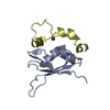
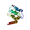
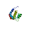
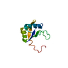
| Title | Crystal structure of de novo designed MID1-cobalt | ||||||
 Components Components | Computational design, MID1-cobalt | ||||||
 Keywords Keywords | DE NOVO PROTEIN / METAL BINDING PROTEIN / Helix-turn-helix / Zinc homodimerization | ||||||
| Function / homology | Rabenosyn, Rab binding domain / DNA Excision Repair, Uvrb; Chain A / Few Secondary Structures / Irregular / :  Function and homology information Function and homology information | ||||||
| Biological species | ARTIFICIAL GENE (others) | ||||||
| Method |  X-RAY DIFFRACTION / SYNCHROTRON / MOLECULAR REPLACEMENT / Resolution: 1.239 Å X-RAY DIFFRACTION / SYNCHROTRON / MOLECULAR REPLACEMENT / Resolution: 1.239 Å | ||||||
 Authors Authors | Der, B.S. / Machius, M. / Miley, M.J. / Kuhlman, B. | ||||||
 Citation Citation | Journal: J.Am.Chem.Soc. / Year: 2012 Title: Metal-mediated affinity and orientation specificity in a computationally designed protein homodimer. Authors: Der, B.S. / Machius, M. / Miley, M.J. / Mills, J.L. / Szyperski, T. / Kuhlman, B. | ||||||
| History |
|
- Structure visualization
| Structure viewer | Molecule: MolmilJmol/JSmol |
|---|
- Downloads & links
-Download
| PDBx/mmCIF format | 3v1d.cif.gz | 261.9 KB | Display | PDBx/mmCIF format |
|---|---|---|---|---|
| PDB format | pdb3v1d.ent.gz | 217.3 KB | Display | PDB format |
| PDBx/mmJSON format | 3v1d.json.gz | Tree view | PDBx/mmJSON format | |
| Others |  Other downloads Other downloads |
-Validation report
| Arichive directory | https://data.pdbj.org/pub/pdb/validation_reports/v1/3v1dftp://data.pdbj.org/pub/pdb/validation_reports/v1/3v1d | HTTPS FTP |
|---|
-Related structure data
| Related structure data |  3v1aC  3v1bC 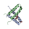 3v1cC  3v1eC  3v1fC  1yzmS S: Starting model for refinement C: citing same article ( |
|---|---|
| Similar structure data |


-Links
 PDBj
PDBj

- Assembly
| Deposited unit | 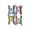
| ||||||||
|---|---|---|---|---|---|---|---|---|---|
| 1 | 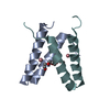
| ||||||||
| 2 | 
| ||||||||
| 3 | 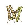
| ||||||||
| 4 | 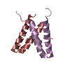
| ||||||||
| Unit cell |
|
-Components
| #1: Protein/peptide | Mass: 5499.118 Da / Num. of mol.: 8 Source method: isolated from a genetically manipulated source Source: (gene. exp.) ARTIFICIAL GENE (others) / Plasmid: pQE-80L MBP fusion / Production host:  #2: Chemical | ChemComp-CO /   Mass: 58.933 Da / Num. of mol.: 8 / Source method: obtained synthetically / Formula: Co Mass: 58.933 Da / Num. of mol.: 8 / Source method: obtained synthetically / Formula: Co#3: Chemical | ChemComp-1PE /   Mass: 238.278 Da / Num. of mol.: 4 / Source method: obtained synthetically / Formula: C10H22O6 / Comment: precipitant*YM Mass: 238.278 Da / Num. of mol.: 4 / Source method: obtained synthetically / Formula: C10H22O6 / Comment: precipitant*YM#4: Water | ChemComp-HOH / |  Mass: 18.015 Da / Num. of mol.: 393 / Source method: isolated from a natural source / Formula: H2O Mass: 18.015 Da / Num. of mol.: 393 / Source method: isolated from a natural source / Formula: H2O |
|---|
-Experimental details
-Experiment
| Experiment | Method: X-RAY DIFFRACTION / Number of used crystals: 1 |
|---|
- Sample preparation
| Crystal | Density Matthews: 1.78 Å3/Da / Density % sol: 30.85 % |
|---|---|
| Crystal grow | Temperature: 293 K / Method: vapor diffusion, hanging drop / pH: 6 Details: 1-2 microliters protein (20 mg/ml, 100 mM ammonium acetate buffer) mixed with 1 microliter crystallization buffer (0.1 M MES pH 6.0, 30% PEG 200, 10% PEG 3000), VAPOR DIFFUSION, HANGING DROP, temperature 293K |
-Data collection
| Diffraction | Mean temperature: 100 K |
|---|---|
| Diffraction source | Source: SYNCHROTRON / Site: APS  / Beamline: 23-ID-B / Wavelength: 0.9494 Å / Beamline: 23-ID-B / Wavelength: 0.9494 Å |
| Detector | Type: MARMOSAIC 300 mm CCD / Detector: CCD / Date: Apr 26, 2011 |
| Radiation | Monochromator: double crystal / Protocol: SINGLE WAVELENGTH / Monochromatic (M) / Laue (L): M / Scattering type: x-ray |
| Radiation wavelength | Wavelength: 0.9494 Å / Relative weight: 1 |
| Reflection | Resolution: 1.239→36.668 Å / Num. all: 81186 / Num. obs: 81186 / % possible obs: 94.3 % / Observed criterion σ(F): -3 / Observed criterion σ(I): -3 / Redundancy: 2.5 % / Biso Wilson estimate: 7.8 Å2 / Rmerge(I) obs: 0.068 / Net I/σ(I): 18.1 |
| Reflection shell | Resolution: 1.24→1.25 Å / Redundancy: 2 % / Rmerge(I) obs: 0.429 / Mean I/σ(I) obs: 2 / % possible all: 82.6 |
- Processing
| Software |
| |||||||||||||||||||||||||||||||||||||||||||||||||||||||||||||||
|---|---|---|---|---|---|---|---|---|---|---|---|---|---|---|---|---|---|---|---|---|---|---|---|---|---|---|---|---|---|---|---|---|---|---|---|---|---|---|---|---|---|---|---|---|---|---|---|---|---|---|---|---|---|---|---|---|---|---|---|---|---|---|---|---|
| Refinement | Method to determine structure: MOLECULAR REPLACEMENT Starting model: PDB ENTRY 1YZM Resolution: 1.239→36.668 Å / SU ML: 0.14 / Isotropic thermal model: Anisotropic / Cross valid method: THROUGHOUT / σ(F): 1.96 / Phase error: 19.58 / Stereochemistry target values: ML
| |||||||||||||||||||||||||||||||||||||||||||||||||||||||||||||||
| Solvent computation | Shrinkage radii: 0.86 Å / VDW probe radii: 1.1 Å / Solvent model: FLAT BULK SOLVENT MODEL / Bsol: 63.972 Å2 / ksol: 0.462 e/Å3 | |||||||||||||||||||||||||||||||||||||||||||||||||||||||||||||||
| Displacement parameters |
| |||||||||||||||||||||||||||||||||||||||||||||||||||||||||||||||
| Refinement step | Cycle: LAST / Resolution: 1.239→36.668 Å
| |||||||||||||||||||||||||||||||||||||||||||||||||||||||||||||||
| Refine LS restraints |
| |||||||||||||||||||||||||||||||||||||||||||||||||||||||||||||||
| LS refinement shell |
|
