 Movie
Movie Controller

[English] 日本語
 Yorodumi
- PDB-5c0k: Fragment-Based Drug Discovery Targeting Inhibitor of Apoptosis Pr... -
+ Open data

- Basic information
| Entry | Database: PDB / ID: 5c0k | ||||||
|---|---|---|---|---|---|---|---|
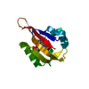
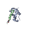
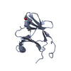
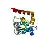
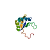
| Title | Fragment-Based Drug Discovery Targeting Inhibitor of Apoptosis Proteins: Compound 3 | ||||||
 Components Components | E3 ubiquitin-protein ligase XIAP | ||||||
 Keywords Keywords | APOPTOSIS / ligase | ||||||
| Function / homology |  Function and homology information Function and homology informationregulation of apoptosis involved in tissue homeostasis / positive regulation of protein linear polyubiquitination / regulation of BMP signaling pathway / nucleotide-binding oligomerization domain containing 1 signaling pathway / copper ion homeostasis / regulation of nucleotide-binding domain, leucine rich repeat containing receptor signaling pathway / nucleotide-binding oligomerization domain containing 2 signaling pathway / SMAC, XIAP-regulated apoptotic response / cysteine-type endopeptidase inhibitor activity involved in apoptotic process / Activation of caspases through apoptosome-mediated cleavage ...regulation of apoptosis involved in tissue homeostasis / positive regulation of protein linear polyubiquitination / regulation of BMP signaling pathway / nucleotide-binding oligomerization domain containing 1 signaling pathway / copper ion homeostasis / regulation of nucleotide-binding domain, leucine rich repeat containing receptor signaling pathway / nucleotide-binding oligomerization domain containing 2 signaling pathway / SMAC, XIAP-regulated apoptotic response / cysteine-type endopeptidase inhibitor activity involved in apoptotic process / Activation of caspases through apoptosome-mediated cleavage / SMAC (DIABLO) binds to IAPs / SMAC(DIABLO)-mediated dissociation of IAP:caspase complexes / Regulation of the apoptosome activity / TNFR1-induced proapoptotic signaling / RIPK1-mediated regulated necrosis / regulation of innate immune response / negative regulation of tumor necrosis factor-mediated signaling pathway / cysteine-type endopeptidase inhibitor activity / protein K63-linked ubiquitination / positive regulation of type I interferon production / positive regulation of protein ubiquitination / Regulation of PTEN localization / protein serine/threonine kinase binding / TNFR1-induced NF-kappa-B signaling pathway / Deactivation of the beta-catenin transactivating complex / Regulation of TNFR1 signaling / RING-type E3 ubiquitin transferase / positive regulation of JNK cascade / Regulation of necroptotic cell death / Wnt signaling pathway / Regulation of PTEN stability and activity / ubiquitin-protein transferase activity / positive regulation of canonical Wnt signaling pathway / ubiquitin protein ligase activity / regulation of inflammatory response / response to lipopolysaccharide / regulation of apoptotic process / positive regulation of canonical NF-kappaB signal transduction / regulation of cell cycle / defense response to bacterium / apoptotic process / DNA damage response / negative regulation of apoptotic process / nucleolus / zinc ion binding / nucleoplasm / identical protein binding / nucleus / cytosol / cytoplasm Similarity search - Function | ||||||
| Biological species |  Homo sapiens (human) Homo sapiens (human) | ||||||
| Method |  X-RAY DIFFRACTION / SYNCHROTRON / MOLECULAR REPLACEMENT / Resolution: 2.2 Å X-RAY DIFFRACTION / SYNCHROTRON / MOLECULAR REPLACEMENT / Resolution: 2.2 Å | ||||||
 Authors Authors | Chessari, G. / Buck, I.M. / Day, J.E.H. / Day, P.J. / Iqbal, A. / Johnson, C.N. / Lewis, E.J. / Martins, V. / Miller, D. / Reader, M. ...Chessari, G. / Buck, I.M. / Day, J.E.H. / Day, P.J. / Iqbal, A. / Johnson, C.N. / Lewis, E.J. / Martins, V. / Miller, D. / Reader, M. / Rees, D.C. / Rich, S.J. / Tamanini, E. / Vitorino, M. / Ward, G.A. / Williams, P.A. / Williams, G. / Wilsher, N.E. / Woolford, A.J.-A. | ||||||
 Citation Citation | Journal: J.Med.Chem. / Year: 2015 Title: Fragment-Based Drug Discovery Targeting Inhibitor of Apoptosis Proteins: Discovery of a Non-Alanine Lead Series with Dual Activity Against cIAP1 and XIAP. Authors: Chessari, G. / Buck, I.M. / Day, J.E. / Day, P.J. / Iqbal, A. / Johnson, C.N. / Lewis, E.J. / Martins, V. / Miller, D. / Reader, M. / Rees, D.C. / Rich, S.J. / Tamanini, E. / Vitorino, M. / ...Authors: Chessari, G. / Buck, I.M. / Day, J.E. / Day, P.J. / Iqbal, A. / Johnson, C.N. / Lewis, E.J. / Martins, V. / Miller, D. / Reader, M. / Rees, D.C. / Rich, S.J. / Tamanini, E. / Vitorino, M. / Ward, G.A. / Williams, P.A. / Williams, G. / Wilsher, N.E. / Woolford, A.J. | ||||||
| History |
|
- Structure visualization
| Structure viewer | Molecule: MolmilJmol/JSmol |
|---|
- Downloads & links
-Download
| PDBx/mmCIF format | 5c0k.cif.gz | 63.1 KB | Display | PDBx/mmCIF format |
|---|---|---|---|---|
| PDB format | pdb5c0k.ent.gz | 45 KB | Display | PDB format |
| PDBx/mmJSON format | 5c0k.json.gz | Tree view | PDBx/mmJSON format | |
| Others |  Other downloads Other downloads |
-Validation report
| Arichive directory | https://data.pdbj.org/pub/pdb/validation_reports/c0/5c0kftp://data.pdbj.org/pub/pdb/validation_reports/c0/5c0k | HTTPS FTP |
|---|
-Related structure data
| Related structure data | 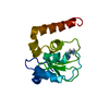 5c0lC 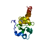 5c3hC 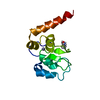 5c3kC 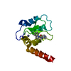 5c7aC 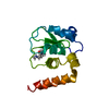 5c7bC 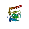 5c7cC 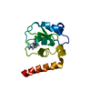 5c7dC 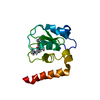 5c83C 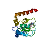 5c84C 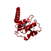 2opyS S: Starting model for refinement C: citing same article ( |
|---|---|
| Similar structure data |


-Links
 PDBj
PDBj


- Assembly
| Deposited unit | 
| |||||||||
|---|---|---|---|---|---|---|---|---|---|---|
| 1 |
| |||||||||
| Unit cell |
| |||||||||
| Components on special symmetry positions |
|
-Components
| #1: Protein | Mass: 12686.206 Da / Num. of mol.: 1 / Fragment: residues 249-354 Source method: isolated from a genetically manipulated source Details: Image clone / Source: (gene. exp.) Homo sapiens (human) / Gene: XIAP, API3, BIRC4, IAP3 / Plasmid: pET 28b / Production host:  References: UniProt: P98170, Ligases; Forming carbon-nitrogen bonds; Acid-amino-acid ligases (peptide synthases) | ||||
|---|---|---|---|---|---|
| #2: Chemical | ChemComp-ZN /   Mass: 65.409 Da / Num. of mol.: 1 / Source method: obtained synthetically / Formula: Zn Mass: 65.409 Da / Num. of mol.: 1 / Source method: obtained synthetically / Formula: Zn | ||||
| #3: Chemical |   Mass: 62.068 Da / Num. of mol.: 2 / Source method: obtained synthetically / Formula: C2H6O2 Mass: 62.068 Da / Num. of mol.: 2 / Source method: obtained synthetically / Formula: C2H6O2#4: Chemical | ChemComp-4WK / ( |   Mass: 188.247 Da / Num. of mol.: 1 / Source method: obtained synthetically / Formula: C8H18N3O2 Mass: 188.247 Da / Num. of mol.: 1 / Source method: obtained synthetically / Formula: C8H18N3O2#5: Water | ChemComp-HOH / |  Mass: 18.015 Da / Num. of mol.: 126 / Source method: isolated from a natural source / Formula: H2O Mass: 18.015 Da / Num. of mol.: 126 / Source method: isolated from a natural source / Formula: H2O |
-Experimental details
-Experiment
| Experiment | Method: X-RAY DIFFRACTION |
|---|
- Sample preparation
| Crystal | Density Matthews: 2.32 Å3/Da / Density meas: 1.35 Mg/m3 / Density % sol: 47 % / Description: Blocks |
|---|---|
| Crystal grow | Temperature: 277 K / Method: vapor diffusion, hanging drop / pH: 7.5 / Details: 3.9M NaCl, 0.1M HEPES/NaOH pH=7.5 / PH range: 7.5 |
-Data collection
| Diffraction | Mean temperature: 100 K |
|---|---|
| Diffraction source | Source: SYNCHROTRON / Site: ESRF  / Beamline: ID23-1 / Wavelength: 0.9 Å / Beamline: ID23-1 / Wavelength: 0.9 Å |
| Detector | Type: ADSC QUANTUM 4 / Detector: CCD / Date: Nov 11, 2008 |
| Radiation | Protocol: SINGLE WAVELENGTH / Monochromatic (M) / Laue (L): M / Scattering type: x-ray |
| Radiation wavelength | Wavelength: 0.9 Å / Relative weight: 1 |
| Reflection | Resolution: 2.2→27.29 Å / Num. all: 14177 / Num. obs: 14177 / % possible obs: 98.5 % / Observed criterion σ(I): 2 / Redundancy: 3.58 % / Biso Wilson estimate: 60.57 Å2 / Rmerge(I) obs: 0.066 / Net I/σ(I): 8.6 |
| Reflection shell | Resolution: 2.2→2.28 Å / Rmerge(I) obs: 0.392 / Mean I/σ(I) obs: 2 / % possible all: 99.2 |
- Processing
| Software |
| ||||||||||||||||||||||||||||||||||||||||||||||||||||||||||||||||||||||||||||||||||||||||||||||||||||||||||||||||||
|---|---|---|---|---|---|---|---|---|---|---|---|---|---|---|---|---|---|---|---|---|---|---|---|---|---|---|---|---|---|---|---|---|---|---|---|---|---|---|---|---|---|---|---|---|---|---|---|---|---|---|---|---|---|---|---|---|---|---|---|---|---|---|---|---|---|---|---|---|---|---|---|---|---|---|---|---|---|---|---|---|---|---|---|---|---|---|---|---|---|---|---|---|---|---|---|---|---|---|---|---|---|---|---|---|---|---|---|---|---|---|---|---|---|---|---|
| Refinement | Method to determine structure: MOLECULAR REPLACEMENT Starting model: 2OPY Resolution: 2.2→27.29 Å / Cor.coef. Fo:Fc: 0.9484 / Cor.coef. Fo:Fc free: 0.9397 / SU R Cruickshank DPI: 0.139 / Cross valid method: THROUGHOUT / σ(F): 0 / SU R Blow DPI: 0.156 / SU Rfree Blow DPI: 0.141 / SU Rfree Cruickshank DPI: 0.131
| ||||||||||||||||||||||||||||||||||||||||||||||||||||||||||||||||||||||||||||||||||||||||||||||||||||||||||||||||||
| Displacement parameters | Biso mean: 62.5 Å2
| ||||||||||||||||||||||||||||||||||||||||||||||||||||||||||||||||||||||||||||||||||||||||||||||||||||||||||||||||||
| Refine analyze | Luzzati coordinate error obs: 0.289 Å | ||||||||||||||||||||||||||||||||||||||||||||||||||||||||||||||||||||||||||||||||||||||||||||||||||||||||||||||||||
| Refinement step | Cycle: LAST / Resolution: 2.2→27.29 Å
| ||||||||||||||||||||||||||||||||||||||||||||||||||||||||||||||||||||||||||||||||||||||||||||||||||||||||||||||||||
| Refine LS restraints |
| ||||||||||||||||||||||||||||||||||||||||||||||||||||||||||||||||||||||||||||||||||||||||||||||||||||||||||||||||||
| LS refinement shell | Resolution: 2.2→2.38 Å / Total num. of bins used: 7
| ||||||||||||||||||||||||||||||||||||||||||||||||||||||||||||||||||||||||||||||||||||||||||||||||||||||||||||||||||
| Refinement TLS params. | Method: refined / Origin x: -17.073 Å / Origin y: -29.2196 Å / Origin z: -4.1285 Å
| ||||||||||||||||||||||||||||||||||||||||||||||||||||||||||||||||||||||||||||||||||||||||||||||||||||||||||||||||||
| Refinement TLS group | Selection details: { A|249 - A|352 } |
