 Movie
Movie Controller

[English] 日本語
 Yorodumi
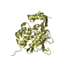
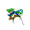
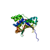
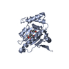
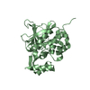
- PDB-3u67: Crystal structure of the N-terminal domain of Hsp90 from Leishman... -
+ Open data

- Basic information
| Entry | Database: PDB / ID: 3u67 | ||||||
|---|---|---|---|---|---|---|---|
| Title | Crystal structure of the N-terminal domain of Hsp90 from Leishmania major(LmjF33.0312)in complex with ADP | ||||||
 Components Components | Heat shock protein 83-1 | ||||||
 Keywords Keywords | CHAPERONE / Structural Genomics / Structural Genomics Consortium / SGC / ATPase / ATP binding | ||||||
| Function / homology |  Function and homology information Function and homology informationATP-dependent protein folding chaperone / : / cellular response to heat / protein folding / protein stabilization / perinuclear region of cytoplasm / ATP hydrolysis activity / protein-containing complex / nucleoplasm / ATP binding ...ATP-dependent protein folding chaperone / : / cellular response to heat / protein folding / protein stabilization / perinuclear region of cytoplasm / ATP hydrolysis activity / protein-containing complex / nucleoplasm / ATP binding / metal ion binding / plasma membrane / cytosol / cytoplasm Similarity search - Function | ||||||
| Biological species |  Leishmania major (eukaryote) Leishmania major (eukaryote) | ||||||
| Method |  X-RAY DIFFRACTION / MOLECULAR REPLACEMENT / Resolution: 1.77 Å X-RAY DIFFRACTION / MOLECULAR REPLACEMENT / Resolution: 1.77 Å | ||||||
 Authors Authors | Pizarro, J.C. / Wernimont, A.K. / Hutchinson, A. / Mackenzie, F. / Fairlamb, A. / Arrowsmith, C.H. / Bountra, C. / Weigelt, J. / Edwards, A.M. / Ferguson, M.A.J. ...Pizarro, J.C. / Wernimont, A.K. / Hutchinson, A. / Mackenzie, F. / Fairlamb, A. / Arrowsmith, C.H. / Bountra, C. / Weigelt, J. / Edwards, A.M. / Ferguson, M.A.J. / Hui, R. / Hills, T. / Structural Genomics Consortium (SGC) | ||||||
 Citation Citation | Journal: PLoS Negl Trop Dis / Year: 2013 Title: Exploring the Trypanosoma brucei Hsp83 potential as a target for structure guided drug design. Authors: Pizarro, J.C. / Hills, T. / Senisterra, G. / Wernimont, A.K. / Mackenzie, C. / Norcross, N.R. / Ferguson, M.A. / Wyatt, P.G. / Gilbert, I.H. / Hui, R. | ||||||
| History |
|
- Structure visualization
| Structure viewer | Molecule: MolmilJmol/JSmol |
|---|
- Downloads & links
-Download
| PDBx/mmCIF format | 3u67.cif.gz | 65.5 KB | Display | PDBx/mmCIF format |
|---|---|---|---|---|
| PDB format | pdb3u67.ent.gz | 45.5 KB | Display | PDB format |
| PDBx/mmJSON format | 3u67.json.gz | Tree view | PDBx/mmJSON format | |
| Others |  Other downloads Other downloads |
-Validation report
| Arichive directory | https://data.pdbj.org/pub/pdb/validation_reports/u6/3u67ftp://data.pdbj.org/pub/pdb/validation_reports/u6/3u67 | HTTPS FTP |
|---|
-Related structure data
| Related structure data | 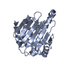 3o6oC 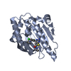 3omuC 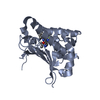 3opdC 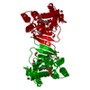 3h80S S: Starting model for refinement C: citing same article ( |
|---|---|
| Similar structure data |


-Links
 PDBj
PDBj

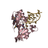
- Assembly
| Deposited unit | 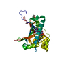
| ||||||||
|---|---|---|---|---|---|---|---|---|---|
| 1 |
| ||||||||
| 2 | 
| ||||||||
| Unit cell |
|
-Components
| #1: Protein | Mass: 26110.465 Da / Num. of mol.: 1 / Fragment: N-terminal domain, residues 1-213 Source method: isolated from a genetically manipulated source Source: (gene. exp.) Leishmania major (eukaryote) / Strain: FRIEDLINGene: HSP83, HSP83-10, HSP83-11, HSP83-12, HSP83-13, HSP83-14, HSP83-15, HSP83-16, HSP83-17, HSP83-2, HSP83-3, HSP83-5, HSP83-6, HSP83-7, HSP83-9, LMJF_33_0312, LMJF_33_0314, LMJF_33_0316, LMJF_33_ ...Gene: HSP83, HSP83-10, HSP83-11, HSP83-12, HSP83-13, HSP83-14, HSP83-15, HSP83-16, HSP83-17, HSP83-2, HSP83-3, HSP83-5, HSP83-6, HSP83-7, HSP83-9, LMJF_33_0312, LMJF_33_0314, LMJF_33_0316, LMJF_33_0320, LMJF_33_0323, LMJF_33_0326, LMJF_33_0333, LMJF_33_0336, LMJF_33_0340, LMJF_33_0343, LMJF_33_0346, LMJF_33_0350, LMJF_33_0355, LMJF_33_0360, LMJF_33_0365 Plasmid: pET15 / Production host:  | ||
|---|---|---|---|
| #2: Chemical | ChemComp-ADP /   Mass: 427.201 Da / Num. of mol.: 1 / Source method: obtained synthetically / Formula: C10H15N5O10P2 / Comment: ADP, energy-carrying molecule*YM Mass: 427.201 Da / Num. of mol.: 1 / Source method: obtained synthetically / Formula: C10H15N5O10P2 / Comment: ADP, energy-carrying molecule*YM | ||
| #3: Chemical | ChemComp-MG /   Mass: 24.305 Da / Num. of mol.: 1 / Source method: obtained synthetically / Formula: Mg Mass: 24.305 Da / Num. of mol.: 1 / Source method: obtained synthetically / Formula: Mg | ||
| #4: Chemical |   Mass: 62.068 Da / Num. of mol.: 3 / Source method: obtained synthetically / Formula: C2H6O2 Mass: 62.068 Da / Num. of mol.: 3 / Source method: obtained synthetically / Formula: C2H6O2#5: Water | ChemComp-HOH / |  Mass: 18.015 Da / Num. of mol.: 187 / Source method: isolated from a natural source / Formula: H2O Mass: 18.015 Da / Num. of mol.: 187 / Source method: isolated from a natural source / Formula: H2O |
-Experimental details
-Experiment
| Experiment | Method: X-RAY DIFFRACTION / Number of used crystals: 1 |
|---|
- Sample preparation
| Crystal | Density Matthews: 2.57 Å3/Da / Density % sol: 52.16 % |
|---|---|
| Crystal grow | Temperature: 291 K / Method: vapor diffusion, sitting drop / pH: 5.5 Details: 25% PEG3350, 0.1M NH4SO4, 0.1M bis-tris, 4.15mM ADP, 5mM MgCl2 , pH 5.5, VAPOR DIFFUSION, SITTING DROP, temperature 291K |
-Data collection
| Diffraction | Mean temperature: 158 K |
|---|---|
| Diffraction source | Source: ROTATING ANODE / Type: RIGAKU MICROMAX-007 / Wavelength: 1.54 Å |
| Detector | Type: RIGAKU RAXIS IV++ / Detector: IMAGE PLATE / Date: Jun 13, 2009 / Details: mirrors |
| Radiation | Monochromator: graphite / Protocol: SINGLE WAVELENGTH / Monochromatic (M) / Laue (L): M / Scattering type: x-ray |
| Radiation wavelength | Wavelength: 1.54 Å / Relative weight: 1 |
| Reflection | Resolution: 1.77→50 Å / Num. obs: 26898 / % possible obs: 99.8 % / Observed criterion σ(F): 0 / Observed criterion σ(I): 0 / Redundancy: 6.7 % / Biso Wilson estimate: 31.55 Å2 / Rsym value: 0.093 / Net I/σ(I): 18.9 |
| Reflection shell | Resolution: 1.77→1.83 Å / Redundancy: 5.8 % / Num. unique all: 2586 / Rsym value: 0.492 / % possible all: 98.4 |
- Processing
| Software |
| ||||||||||||||||||||||||||||||||||||||||||||||||||||||||||||||||||||||||||||||||||||||||||||||||||||||||||||||||||
|---|---|---|---|---|---|---|---|---|---|---|---|---|---|---|---|---|---|---|---|---|---|---|---|---|---|---|---|---|---|---|---|---|---|---|---|---|---|---|---|---|---|---|---|---|---|---|---|---|---|---|---|---|---|---|---|---|---|---|---|---|---|---|---|---|---|---|---|---|---|---|---|---|---|---|---|---|---|---|---|---|---|---|---|---|---|---|---|---|---|---|---|---|---|---|---|---|---|---|---|---|---|---|---|---|---|---|---|---|---|---|---|---|---|---|---|
| Refinement | Method to determine structure: MOLECULAR REPLACEMENT Starting model: pdb entry 3H80 Resolution: 1.77→32.94 Å / Cor.coef. Fo:Fc: 0.9293 / Cor.coef. Fo:Fc free: 0.9177 / Cross valid method: THROUGHOUT / σ(F): 0 / Stereochemistry target values: Engh & Huber
| ||||||||||||||||||||||||||||||||||||||||||||||||||||||||||||||||||||||||||||||||||||||||||||||||||||||||||||||||||
| Displacement parameters | Biso mean: 36.73 Å2
| ||||||||||||||||||||||||||||||||||||||||||||||||||||||||||||||||||||||||||||||||||||||||||||||||||||||||||||||||||
| Refine analyze | Luzzati coordinate error obs: 0.217 Å | ||||||||||||||||||||||||||||||||||||||||||||||||||||||||||||||||||||||||||||||||||||||||||||||||||||||||||||||||||
| Refinement step | Cycle: LAST / Resolution: 1.77→32.94 Å
| ||||||||||||||||||||||||||||||||||||||||||||||||||||||||||||||||||||||||||||||||||||||||||||||||||||||||||||||||||
| Refine LS restraints |
| ||||||||||||||||||||||||||||||||||||||||||||||||||||||||||||||||||||||||||||||||||||||||||||||||||||||||||||||||||
| LS refinement shell | Resolution: 1.77→1.84 Å / Total num. of bins used: 13
|
