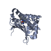
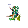
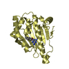
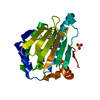
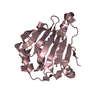
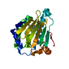
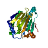
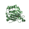
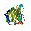
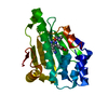
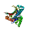
ATP-dependent protein folding chaperone / : / cellular response to heat / protein folding / protein stabilization / perinuclear region of cytoplasm / ATP hydrolysis activity / protein-containing complex / ATP binding / plasma membrane / cytosol Similarity search - Function
Heat shock protein Hsp90, conserved site / Heat shock hsp90 proteins family signature. / Histidine kinase-like ATPase, C-terminal domain / Heat Shock Protein 90 / HSP90, C-terminal domain / Heat shock protein Hsp90, N-terminal / Heat shock protein Hsp90 family / Hsp90 protein / Histidine kinase-, DNA gyrase B-, and HSP90-like ATPase / Histidine kinase-like ATPases ...Heat shock protein Hsp90, conserved site / Heat shock hsp90 proteins family signature. / Histidine kinase-like ATPase, C-terminal domain / Heat Shock Protein 90 / HSP90, C-terminal domain / Heat shock protein Hsp90, N-terminal / Heat shock protein Hsp90 family / Hsp90 protein / Histidine kinase-, DNA gyrase B-, and HSP90-like ATPase / Histidine kinase-like ATPases / Histidine kinase/HSP90-like ATPase / Histidine kinase/HSP90-like ATPase superfamily / Ribosomal protein S5 domain 2-type fold / 2-Layer Sandwich / Alpha Beta Similarity search - Domain/homology
Mass: 18.015 Da / Num. of mol.: 96 / Source method: isolated from a natural source / Formula: H2O
Has protein modification
Y
-
Experimental details
-
Experiment
Experiment
Method: X-RAY DIFFRACTION / Number of used crystals: 1
-
Sample preparation
Crystal
Density Matthews: 2.22 Å3/Da / Density % sol: 44.52 %
Crystal grow
Temperature: 293 K / Method: vapor diffusion, sitting drop / pH: 7.5 Details: 25% PEG 3350 0.2 M Ammonium Acetate 0.1 M Hepes pH 7.5 4 mM MgCl2 2 mM TCEP 2 mM DDU101329, VAPOR DIFFUSION, SITTING DROP, temperature 293K
Resolution: 2.15→28.16 Å / Cor.coef. Fo:Fc: 0.937 / Cor.coef. Fo:Fc free: 0.902 / WRfactor Rfree: 0.2619 / WRfactor Rwork: 0.2147 / Occupancy max: 1 / Occupancy min: 0.5 / FOM work R set: 0.8281 / SU B: 12.683 / SU ML: 0.162 / SU R Cruickshank DPI: 0.2737 / SU Rfree: 0.2235 / Cross valid method: THROUGHOUT / σ(F): 0 / ESU R Free: 0.224 / Stereochemistry target values: MAXIMUM LIKELIHOOD Details: HYDROGENS HAVE BEEN ADDED IN THE RIDING POSITIONS U VALUES : RESIDUAL ONLY
Rfactor
Num. reflection
% reflection
Selection details
Rfree
0.273
1298
5.1 %
RANDOM
Rwork
0.2239
-
-
-
obs
0.2264
25447
97.27 %
-
all
-
26160
-
-
Solvent computation
Ion probe radii: 0.8 Å / Shrinkage radii: 0.8 Å / VDW probe radii: 1.4 Å / Solvent model: MASK
In the structure databanks used in Yorodumi, some data are registered as the other names, "COVID-19 virus" and "2019-nCoV". Here are the details of the virus and the list of structure data.
Jan 31, 2019. EMDB accession codes are about to change! (news from PDBe EMDB page)
EMDB accession codes are about to change! (news from PDBe EMDB page)
The allocation of 4 digits for EMDB accession codes will soon come to an end. Whilst these codes will remain in use, new EMDB accession codes will include an additional digit and will expand incrementally as the available range of codes is exhausted. The current 4-digit format prefixed with “EMD-” (i.e. EMD-XXXX) will advance to a 5-digit format (i.e. EMD-XXXXX), and so on. It is currently estimated that the 4-digit codes will be depleted around Spring 2019, at which point the 5-digit format will come into force.
The EM Navigator/Yorodumi systems omit the EMD- prefix.
Related info.:Q: What is EMD? / ID/Accession-code notation in Yorodumi/EM Navigator
Yorodumi is a browser for structure data from EMDB, PDB, SASBDB, etc.
This page is also the successor to EM Navigator detail page, and also detail information page/front-end page for Omokage search.
The word "yorodu" (or yorozu) is an old Japanese word meaning "ten thousand". "mi" (miru) is to see.
Related info.:EMDB / PDB / SASBDB / Comparison of 3 databanks / Yorodumi Search / Aug 31, 2016. New EM Navigator & Yorodumi / Yorodumi Papers / Jmol/JSmol / Function and homology information / Changes in new EM Navigator and Yorodumi
 Movie
Movie Controller

 Yorodumi
 Open data
 Basic information
 Components
 Keywords
 Function and homology information
 X-RAY DIFFRACTION /
  Authors
 Citation
 Structure visualization
 Downloads & links
 Other downloads
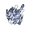

 PDBj
PDBj

 Assembly
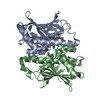


 Mass: 482.427 Da / Num. of mol.: 2 / Source method: obtained synthetically / Formula: C21H25Cl2N5O2S
 Mass: 18.015 Da / Num. of mol.: 96 / Source method: isolated from a natural source / Formula: H2O
 Sample preparation
 Processing