 Movie
Movie Controller

[English] 日本語
 Yorodumi
- PDB-2i82: Crystal structure of pseudouridine synthase RluA: indirect sequen... -
+ Open data

- Basic information
| Entry | Database: PDB / ID: 2i82 | |||||||||
|---|---|---|---|---|---|---|---|---|---|---|
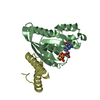
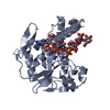
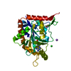
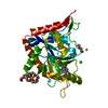
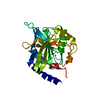
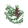
| Title | Crystal structure of pseudouridine synthase RluA: indirect sequence readout through protein-induced RNA structure | |||||||||
 Components Components |
| |||||||||
 Keywords Keywords | Lyase/RNA / pseudouridine synthase / Lyase-RNA COMPLEX | |||||||||
| Function / homology |  Function and homology information Function and homology informationtRNA pseudouridine32 synthase / 23S rRNA pseudouridine746 synthase / 23S rRNA pseudouridine(746) synthase activity / tRNA pseudouridine(32) synthase activity / tRNA pseudouridine synthase activity / rRNA pseudouridine synthase activity / RNA modification / enzyme-directed rRNA pseudouridine synthesis / pseudouridine synthesis / rRNA pseudouridine synthesis ...tRNA pseudouridine32 synthase / 23S rRNA pseudouridine746 synthase / 23S rRNA pseudouridine(746) synthase activity / tRNA pseudouridine(32) synthase activity / tRNA pseudouridine synthase activity / rRNA pseudouridine synthase activity / RNA modification / enzyme-directed rRNA pseudouridine synthesis / pseudouridine synthesis / rRNA pseudouridine synthesis / tRNA pseudouridine synthesis / pseudouridine synthase activity / tRNA processing / rRNA processing / RNA binding / cytosol Similarity search - Function | |||||||||
| Biological species |  | |||||||||
| Method |  X-RAY DIFFRACTION / SYNCHROTRON / MAD / Resolution: 2.05 Å X-RAY DIFFRACTION / SYNCHROTRON / MAD / Resolution: 2.05 Å | |||||||||
 Authors Authors | Hoang, C. | |||||||||
 Citation Citation | Journal: Mol.Cell / Year: 2006 Title: Crystal structure of pseudouridine synthase RluA: indirect sequence readout through protein-induced RNA structure Authors: Hoang, C. / Chen, J. / Vizthum, C.A. / Kandel, J.M. / Hamilton, C.S. / Mueller, E.G. / Ferre-D'Amare, A.R. | |||||||||
| History |
|
- Structure visualization
| Structure viewer | Molecule: MolmilJmol/JSmol |
|---|
- Downloads & links
-Download
| PDBx/mmCIF format | 2i82.cif.gz | 237.7 KB | Display | PDBx/mmCIF format |
|---|---|---|---|---|
| PDB format | pdb2i82.ent.gz | 183.8 KB | Display | PDB format |
| PDBx/mmJSON format | 2i82.json.gz | Tree view | PDBx/mmJSON format | |
| Others |  Other downloads Other downloads |
-Validation report
| Arichive directory | https://data.pdbj.org/pub/pdb/validation_reports/i8/2i82ftp://data.pdbj.org/pub/pdb/validation_reports/i8/2i82 | HTTPS FTP |
|---|
-Related structure data
| Similar structure data |
|---|


-Links
 PDBj
PDBj


- Assembly
| Deposited unit | 
| ||||||||
|---|---|---|---|---|---|---|---|---|---|
| 1 | 
| ||||||||
| 2 | 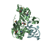
| ||||||||
| 3 | 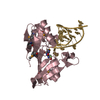
| ||||||||
| 4 | 
| ||||||||
| Unit cell |
| ||||||||
| Details | The crystallographic model consists of all residues for the four RNA molecules in the asymmetric unit. |
-Components
| #1: RNA chain | Mass: 6642.013 Da / Num. of mol.: 4 / Source method: obtained synthetically #2: Protein | Mass: 25083.588 Da / Num. of mol.: 4 Source method: isolated from a genetically manipulated source Source: (gene. exp.) #3: Chemical |   Mass: 150.108 Da / Num. of mol.: 2 / Source method: obtained synthetically / Formula: C4H7FN2O3 Mass: 150.108 Da / Num. of mol.: 2 / Source method: obtained synthetically / Formula: C4H7FN2O3#4: Water | ChemComp-HOH / |  Mass: 18.015 Da / Num. of mol.: 255 / Source method: isolated from a natural source / Formula: H2O Mass: 18.015 Da / Num. of mol.: 255 / Source method: isolated from a natural source / Formula: H2OHas protein modification | Y | |
|---|
-Experimental details
-Experiment
| Experiment | Method: X-RAY DIFFRACTION / Number of used crystals: 2 |
|---|
- Sample preparation
| Crystal | Density Matthews: 2.64 Å3/Da / Density % sol: 53.45 % | ||||||||||||||||||||||||||||||||||||||||||||||||
|---|---|---|---|---|---|---|---|---|---|---|---|---|---|---|---|---|---|---|---|---|---|---|---|---|---|---|---|---|---|---|---|---|---|---|---|---|---|---|---|---|---|---|---|---|---|---|---|---|---|
| Crystal grow | Temperature: 295 K / Method: vapor diffusion, hanging drop / pH: 8.5 Details: 25% PEG4000, 100mM Tris-HCl pH 8.5, 25mM MgCl2, 100mM LiCl, 1mM spermine, VAPOR DIFFUSION, HANGING DROP, temperature 295K | ||||||||||||||||||||||||||||||||||||||||||||||||
| Components of the solutions |
|
-Data collection
| Diffraction | Mean temperature: 100 K | |||||||||||||||||||||
|---|---|---|---|---|---|---|---|---|---|---|---|---|---|---|---|---|---|---|---|---|---|---|
| Diffraction source | Source: SYNCHROTRON / Site: ALS  / Beamline: 8.2.1 / Beamline: 8.2.1Wavelength: 0.9001, 0.92035, 0.92014, 0.942411, 0.979877, 0.979647 | |||||||||||||||||||||
| Detector | Type: ADSC QUANTUM 210 / Detector: CCD / Date: Jul 10, 2005 | |||||||||||||||||||||
| Radiation | Monochromator: Double crystal, Si (111) / Protocol: MAD / Monochromatic (M) / Laue (L): M / Scattering type: x-ray | |||||||||||||||||||||
| Radiation wavelength |
| |||||||||||||||||||||
| Reflection | Resolution: 2.05→19.83 Å / Num. obs: 82366 / % possible obs: 97.8 % / Observed criterion σ(F): 0 / Observed criterion σ(I): 4.84 / Redundancy: 7.43 % / Biso Wilson estimate: 23.1 Å2 / Rmerge(I) obs: 0.069 / Rsym value: 0.069 / Net I/σ(I): 35.8 | |||||||||||||||||||||
| Reflection shell | Resolution: 2.05→2.18 Å / Redundancy: 6.96 % / Rmerge(I) obs: 0.467 / Mean I/σ(I) obs: 4.84 / Num. unique all: 8090 / Rsym value: 0.467 / % possible all: 97.2 |
- Processing
| Software |
| ||||||||||||||||||||||||||||||||||||
|---|---|---|---|---|---|---|---|---|---|---|---|---|---|---|---|---|---|---|---|---|---|---|---|---|---|---|---|---|---|---|---|---|---|---|---|---|---|
| Refinement | Method to determine structure: MAD / Resolution: 2.05→19.83 Å / Rfactor Rfree error: 0.003 / Data cutoff high absF: 2402292.05 / Data cutoff low absF: 0 / Isotropic thermal model: RESTRAINED / Cross valid method: THROUGHOUT / σ(F): 0 / Stereochemistry target values: Engh & Huber
| ||||||||||||||||||||||||||||||||||||
| Solvent computation | Solvent model: FLAT MODEL / Bsol: 47.5999 Å2 / ksol: 0.344821 e/Å3 | ||||||||||||||||||||||||||||||||||||
| Displacement parameters | Biso mean: 41.8 Å2
| ||||||||||||||||||||||||||||||||||||
| Refine analyze |
| ||||||||||||||||||||||||||||||||||||
| Refinement step | Cycle: LAST / Resolution: 2.05→19.83 Å
| ||||||||||||||||||||||||||||||||||||
| Refine LS restraints |
| ||||||||||||||||||||||||||||||||||||
| LS refinement shell | Resolution: 2.05→2.18 Å / Rfactor Rfree error: 0.009 / Total num. of bins used: 6
| ||||||||||||||||||||||||||||||||||||
| Xplor file |
|
