 Movie
Movie Controller

[English] 日本語
 Yorodumi
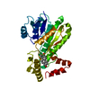
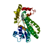
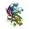
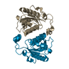
- PDB-3to0: Crystal structure of Mus musculus iodotyrosine deiodinase (IYD) C... -
+ Open data

- Basic information
| Entry | Database: PDB / ID: 3to0 | ||||||
|---|---|---|---|---|---|---|---|
| Title | Crystal structure of Mus musculus iodotyrosine deiodinase (IYD) C217A, C239A bound to FMN | ||||||
 Components Components | Iodotyrosine deiodinase 1 | ||||||
 Keywords Keywords | OXIDOREDUCTASE / FLAVOPROTEIN / MEMBRANE / TRANSMEMBRANE / DEHALOGENASE / IODIDE SALVAGE / MONO-IODOTYROSINE / NADP | ||||||
| Function / homology |  Function and homology information Function and homology informationThyroxine biosynthesis / iodotyrosine deiodinase / iodotyrosine deiodinase activity / : / thyroid hormone metabolic process / cytoplasmic vesicle membrane / FMN binding / oxidoreductase activity / nucleoplasm / plasma membrane Similarity search - Function | ||||||
| Biological species |  | ||||||
| Method |  X-RAY DIFFRACTION / MOLECULAR REPLACEMENT / Resolution: 2.655 Å X-RAY DIFFRACTION / MOLECULAR REPLACEMENT / Resolution: 2.655 Å | ||||||
 Authors Authors | Buss, J.M. / McTamney, P.M. / Rokita, S.E. | ||||||
 Citation Citation | Journal: Protein Sci. / Year: 2012 Title: Expression of a soluble form of iodotyrosine deiodinase for active site characterization by engineering the native membrane protein from Mus musculus. Authors: Buss, J.M. / McTamney, P.M. / Rokita, S.E. | ||||||
| History |
|
- Structure visualization
| Structure viewer | Molecule: MolmilJmol/JSmol |
|---|
- Downloads & links
-Download
| PDBx/mmCIF format | 3to0.cif.gz | 93 KB | Display | PDBx/mmCIF format |
|---|---|---|---|---|
| PDB format | pdb3to0.ent.gz | 69 KB | Display | PDB format |
| PDBx/mmJSON format | 3to0.json.gz | Tree view | PDBx/mmJSON format | |
| Others |  Other downloads Other downloads |
-Validation report
| Arichive directory | https://data.pdbj.org/pub/pdb/validation_reports/to/3to0ftp://data.pdbj.org/pub/pdb/validation_reports/to/3to0 | HTTPS FTP |
|---|
-Related structure data
| Related structure data |  3tnzC 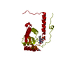 3gb5S S: Starting model for refinement C: citing same article ( |
|---|---|
| Similar structure data |


-Links
 PDBj
PDBj
- Assembly
| Deposited unit | 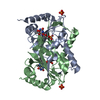
| ||||||||||||||||||
|---|---|---|---|---|---|---|---|---|---|---|---|---|---|---|---|---|---|---|---|
| 1 |
| ||||||||||||||||||
| Unit cell |
| ||||||||||||||||||
| Noncrystallographic symmetry (NCS) | NCS domain:
NCS domain segments:
|
-Components
| #1: Protein | Mass: 29923.273 Da / Num. of mol.: 2 / Mutation: C217A, C239A Source method: isolated from a genetically manipulated source Source: (gene. exp.)  #2: Chemical |   Mass: 456.344 Da / Num. of mol.: 2 / Source method: obtained synthetically / Formula: C17H21N4O9P Mass: 456.344 Da / Num. of mol.: 2 / Source method: obtained synthetically / Formula: C17H21N4O9P#3: Chemical |   Mass: 94.971 Da / Num. of mol.: 2 / Source method: obtained synthetically / Formula: PO4 Mass: 94.971 Da / Num. of mol.: 2 / Source method: obtained synthetically / Formula: PO4#4: Chemical |   Mass: 59.044 Da / Num. of mol.: 2 / Source method: obtained synthetically / Formula: C2H3O2 Mass: 59.044 Da / Num. of mol.: 2 / Source method: obtained synthetically / Formula: C2H3O2#5: Water | ChemComp-HOH / |  Mass: 18.015 Da / Num. of mol.: 50 / Source method: isolated from a natural source / Formula: H2O Mass: 18.015 Da / Num. of mol.: 50 / Source method: isolated from a natural source / Formula: H2O |
|---|
-Experimental details
-Experiment
| Experiment | Method: X-RAY DIFFRACTION / Number of used crystals: 1 |
|---|
- Sample preparation
| Crystal | Density Matthews: 2.3 Å3/Da / Density % sol: 46.62 % |
|---|---|
| Crystal grow | Temperature: 293 K / Method: vapor diffusion, hanging drop / pH: 6 Details: 0.2M ammonium acetate 0.1M Bis-Tris pH 6.0, 45% v/v 2-methyl-2,4-pentanediol, VAPOR DIFFUSION, HANGING DROP, temperature 293K |
-Data collection
| Diffraction | Mean temperature: 100 K |
|---|---|
| Diffraction source | Source: ROTATING ANODE / Type: BRUKER AXS MICROSTAR / Wavelength: 1.54 Å |
| Detector | Type: Bruker Platinum 135 / Detector: CCD / Date: Mar 10, 2010 |
| Radiation | Protocol: SINGLE WAVELENGTH / Monochromatic (M) / Laue (L): M / Scattering type: x-ray |
| Radiation wavelength | Wavelength: 1.54 Å / Relative weight: 1 |
| Reflection | Resolution: 2.655→19.88 Å / Num. obs: 15404 / % possible obs: 99.71 % / Redundancy: 3.7 % / Rsym value: 0.105 / Net I/σ(I): 73 |
| Reflection shell | Resolution: 2.656→2.75 Å / Redundancy: 3.7 % / Mean I/σ(I) obs: 0.81 / Num. unique all: 1493 / Rsym value: 0.534 / % possible all: 80.14 |
- Processing
| Software |
| |||||||||||||||||||||||||||||||||||||||||||||||||||||||||||||||||||||||||||||
|---|---|---|---|---|---|---|---|---|---|---|---|---|---|---|---|---|---|---|---|---|---|---|---|---|---|---|---|---|---|---|---|---|---|---|---|---|---|---|---|---|---|---|---|---|---|---|---|---|---|---|---|---|---|---|---|---|---|---|---|---|---|---|---|---|---|---|---|---|---|---|---|---|---|---|---|---|---|---|
| Refinement | Method to determine structure: MOLECULAR REPLACEMENT Starting model: PDB ENTRY 3GB5 Resolution: 2.655→19.88 Å / SU ML: 0.95 / σ(F): 1.96 / Phase error: 22.04 / Stereochemistry target values: ML
| |||||||||||||||||||||||||||||||||||||||||||||||||||||||||||||||||||||||||||||
| Solvent computation | Shrinkage radii: 0.95 Å / VDW probe radii: 1.2 Å / Solvent model: FLAT BULK SOLVENT MODEL / Bsol: 31.424 Å2 / ksol: 0.394 e/Å3 | |||||||||||||||||||||||||||||||||||||||||||||||||||||||||||||||||||||||||||||
| Displacement parameters |
| |||||||||||||||||||||||||||||||||||||||||||||||||||||||||||||||||||||||||||||
| Refinement step | Cycle: LAST / Resolution: 2.655→19.88 Å
| |||||||||||||||||||||||||||||||||||||||||||||||||||||||||||||||||||||||||||||
| Refine LS restraints |
| |||||||||||||||||||||||||||||||||||||||||||||||||||||||||||||||||||||||||||||
| Refine LS restraints NCS |
| |||||||||||||||||||||||||||||||||||||||||||||||||||||||||||||||||||||||||||||
| LS refinement shell |
| |||||||||||||||||||||||||||||||||||||||||||||||||||||||||||||||||||||||||||||
| Refinement TLS params. | T13: 0.0027 Å2 / Method: refined / Refine-ID: X-RAY DIFFRACTION
| |||||||||||||||||||||||||||||||||||||||||||||||||||||||||||||||||||||||||||||
| Refinement TLS group |
|
