 Movie
Movie Controller

[English] 日本語
 Yorodumi
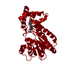
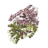
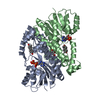
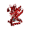
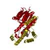
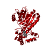
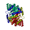
- PDB-1jtv: Crystal structure of 17beta-Hydroxysteroid Dehydrogenase Type 1 c... -
+ Open data

- Basic information
| Entry | Database: PDB / ID: 1jtv | ||||||
|---|---|---|---|---|---|---|---|
| Title | Crystal structure of 17beta-Hydroxysteroid Dehydrogenase Type 1 complexed with Testosterone | ||||||
 Components Components | 17 beta-hydroxysteroid dehydrogenase type 1 | ||||||
 Keywords Keywords | OXIDOREDUCTASE / steroid hormones / alternative binding mode | ||||||
| Function / homology |  Function and homology information Function and homology information17-beta-hydroxysteroid dehydrogenase (NADP+) activity / cellular response to metal ion / estrogen biosynthetic process / 3(or 17)beta-hydroxysteroid dehydrogenase / estradiol binding / Estrogen biosynthesis / testosterone dehydrogenase (NADP+) activity / testosterone biosynthetic process / testosterone dehydrogenase (NAD+) activity / 17beta-estradiol 17-dehydrogenase ...17-beta-hydroxysteroid dehydrogenase (NADP+) activity / cellular response to metal ion / estrogen biosynthetic process / 3(or 17)beta-hydroxysteroid dehydrogenase / estradiol binding / Estrogen biosynthesis / testosterone dehydrogenase (NADP+) activity / testosterone biosynthetic process / testosterone dehydrogenase (NAD+) activity / 17beta-estradiol 17-dehydrogenase / estradiol 17-beta-dehydrogenase [NAD(P)+] activity / NADP+ binding / lysosome organization / The canonical retinoid cycle in rods (twilight vision) / small molecule binding / adipose tissue development / skeletal muscle tissue development / steroid binding / bone development / NADP binding / gene expression / protein homodimerization activity / cytosol Similarity search - Function | ||||||
| Biological species |  Homo sapiens (human) Homo sapiens (human) | ||||||
| Method |  X-RAY DIFFRACTION / SYNCHROTRON / FOURIER SYNTHESIS / Resolution: 1.54 Å X-RAY DIFFRACTION / SYNCHROTRON / FOURIER SYNTHESIS / Resolution: 1.54 Å | ||||||
 Authors Authors | Shi, R. / Nahoum, V. / Lin, S.X. | ||||||
 Citation Citation | Journal: FASEB J. / Year: 2003 Title: Pseudo-symmetry of C19 steroids, alternative binding orientations, and multispecificity in human estrogenic 17beta-hydroxysteroid dehydrogenase. Authors: Gangloff, A. / Shi, R. / Nahoum, V. / Lin, S.X. | ||||||
| History |
|
- Structure visualization
| Structure viewer | Molecule: MolmilJmol/JSmol |
|---|
- Downloads & links
-Download
| PDBx/mmCIF format | 1jtv.cif.gz | 72.5 KB | Display | PDBx/mmCIF format |
|---|---|---|---|---|
| PDB format | pdb1jtv.ent.gz | 51.9 KB | Display | PDB format |
| PDBx/mmJSON format | 1jtv.json.gz | Tree view | PDBx/mmJSON format | |
| Others |  Other downloads Other downloads |
-Validation report
| Arichive directory | https://data.pdbj.org/pub/pdb/validation_reports/jt/1jtvftp://data.pdbj.org/pub/pdb/validation_reports/jt/1jtv | HTTPS FTP |
|---|
-Related structure data
| Related structure data | 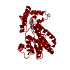 1iolS S: Starting model for refinement |
|---|---|
| Similar structure data |

-Links
 PDBj
PDBj


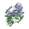
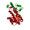
- Assembly
| Deposited unit | 
| ||||||||
|---|---|---|---|---|---|---|---|---|---|
| 1 | 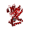
| ||||||||
| Unit cell |
| ||||||||
| Details | The second part of the biological assembly is generated by the two fold crystallographic axis |
-Components
| #1: Protein | Mass: 34887.828 Da / Num. of mol.: 1 / Source method: isolated from a natural source / Source: (natural) Homo sapiens (human) / Tissue: placentaReferences: UniProt: P14061, 17beta-estradiol 17-dehydrogenase |
|---|---|
| #2: Chemical | ChemComp-TES / 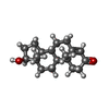  Mass: 288.424 Da / Num. of mol.: 1 / Source method: obtained synthetically / Formula: C19H28O2 / Comment: hormone*YM Mass: 288.424 Da / Num. of mol.: 1 / Source method: obtained synthetically / Formula: C19H28O2 / Comment: hormone*YM |
| #3: Chemical | ChemComp-GOL /   Mass: 92.094 Da / Num. of mol.: 1 / Source method: obtained synthetically / Formula: C3H8O3 Mass: 92.094 Da / Num. of mol.: 1 / Source method: obtained synthetically / Formula: C3H8O3 |
| #4: Water | ChemComp-HOH /  Mass: 18.015 Da / Num. of mol.: 225 / Source method: isolated from a natural source / Formula: H2O Mass: 18.015 Da / Num. of mol.: 225 / Source method: isolated from a natural source / Formula: H2O |
-Experimental details
-Experiment
| Experiment | Method: X-RAY DIFFRACTION / Number of used crystals: 1 |
|---|
- Sample preparation
| Crystal | Density Matthews: 2.3 Å3/Da / Density % sol: 46.61 % | |||||||||||||||||||||||||||||||||||||||||||||||||||||||||||||||||||||||||||||
|---|---|---|---|---|---|---|---|---|---|---|---|---|---|---|---|---|---|---|---|---|---|---|---|---|---|---|---|---|---|---|---|---|---|---|---|---|---|---|---|---|---|---|---|---|---|---|---|---|---|---|---|---|---|---|---|---|---|---|---|---|---|---|---|---|---|---|---|---|---|---|---|---|---|---|---|---|---|---|
| Crystal grow | Temperature: 300 K / pH: 7.5 Details: PEG 4000,magnesium chloride,beta-octylglucoside,glycerol,Hepes,soaking with testosterone, pH 7.5, temperature 300K | |||||||||||||||||||||||||||||||||||||||||||||||||||||||||||||||||||||||||||||
| Crystal grow | *PLUS Temperature: 27 ℃ / Method: vapor diffusion, hanging drop | |||||||||||||||||||||||||||||||||||||||||||||||||||||||||||||||||||||||||||||
| Components of the solutions | *PLUS
|
-Data collection
| Diffraction | Mean temperature: 100 K |
|---|---|
| Diffraction source | Source: SYNCHROTRON / Site: NSLS  / Beamline: X8C / Wavelength: 0.9 Å / Beamline: X8C / Wavelength: 0.9 Å |
| Detector | Type: ADSC QUANTUM 4 / Detector: CCD / Date: Jun 28, 2000 / Details: mirrors |
| Radiation | Protocol: SINGLE WAVELENGTH / Monochromatic (M) / Laue (L): M / Scattering type: x-ray |
| Radiation wavelength | Wavelength: 0.9 Å / Relative weight: 1 |
| Reflection | Resolution: 1.54→40 Å / Num. all: 148865 / Num. obs: 46122 / % possible obs: 98.8 % / Observed criterion σ(I): 1 / Redundancy: 3.2 % / Biso Wilson estimate: 19.4 Å2 / Rmerge(I) obs: 0.049 / Net I/σ(I): 19.1 |
| Reflection shell | Resolution: 1.54→1.6 Å / Redundancy: 3 % / Rmerge(I) obs: 0.42 / Mean I/σ(I) obs: 2 / Num. unique all: 4586 / % possible all: 99.4 |
| Reflection | *PLUS Lowest resolution: 40 Å / Num. measured all: 148865 |
| Reflection shell | *PLUS % possible obs: 99.4 % / Num. unique obs: 4586 / Num. measured obs: 13768 / Rmerge(I) obs: 0.42 / Mean I/σ(I) obs: 2 |
- Processing
| Software |
| |||||||||||||||||||||||||
|---|---|---|---|---|---|---|---|---|---|---|---|---|---|---|---|---|---|---|---|---|---|---|---|---|---|---|
| Refinement | Method to determine structure: FOURIER SYNTHESIS Starting model: PDB ENTRY 1IOL Resolution: 1.54→13.9 Å / Isotropic thermal model: isotropic / Cross valid method: THROUGHOUT / σ(I): 1 / Stereochemistry target values: Engh & Huber
| |||||||||||||||||||||||||
| Displacement parameters | Biso mean: 26.4 Å2 | |||||||||||||||||||||||||
| Refinement step | Cycle: LAST / Resolution: 1.54→13.9 Å
| |||||||||||||||||||||||||
| Refine LS restraints |
| |||||||||||||||||||||||||
| Refinement | *PLUS Lowest resolution: 40 Å | |||||||||||||||||||||||||
| Solvent computation | *PLUS | |||||||||||||||||||||||||
| Displacement parameters | *PLUS | |||||||||||||||||||||||||
| LS refinement shell | *PLUS Highest resolution: 1.54 Å / Lowest resolution: 1.6 Å / Rfactor Rfree: 0.297 / Rfactor Rwork: 0.284 |
