 Movie
Movie Controller

[English] 日本語
 Yorodumi
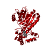
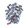
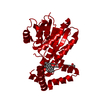
- PDB-1fdw: HUMAN 17-BETA-HYDROXYSTEROID-DEHYDROGENASE TYPE 1 MUTANT H221Q CO... -
+ Open data

- Basic information
| Entry | Database: PDB / ID: 1fdw | ||||||
|---|---|---|---|---|---|---|---|
| Title | HUMAN 17-BETA-HYDROXYSTEROID-DEHYDROGENASE TYPE 1 MUTANT H221Q COMPLEXED WITH ESTRADIOL | ||||||
 Components Components | 17-BETA-HYDROXYSTEROID DEHYDROGENASE | ||||||
 Keywords Keywords | DEHYDROGENASE / 17-BETA-HYDROXYSTEROID / MUTANT / ESTRADIOL / NADP | ||||||
| Function / homology |  Function and homology information Function and homology information17-beta-hydroxysteroid dehydrogenase (NADP+) activity / cellular response to metal ion / estrogen biosynthetic process / 3(or 17)beta-hydroxysteroid dehydrogenase / estradiol binding / Estrogen biosynthesis / testosterone biosynthetic process / testosterone dehydrogenase (NADP+) activity / testosterone dehydrogenase (NAD+) activity / 17beta-estradiol 17-dehydrogenase ...17-beta-hydroxysteroid dehydrogenase (NADP+) activity / cellular response to metal ion / estrogen biosynthetic process / 3(or 17)beta-hydroxysteroid dehydrogenase / estradiol binding / Estrogen biosynthesis / testosterone biosynthetic process / testosterone dehydrogenase (NADP+) activity / testosterone dehydrogenase (NAD+) activity / 17beta-estradiol 17-dehydrogenase / estradiol 17-beta-dehydrogenase [NAD(P)+] activity / NADP+ binding / The canonical retinoid cycle in rods (twilight vision) / small molecule binding / steroid binding / bone development / NADP binding / protein homodimerization activity / cytosol Similarity search - Function | ||||||
| Biological species |  Homo sapiens (human) Homo sapiens (human) | ||||||
| Method |  X-RAY DIFFRACTION / MOLECULAR REPLACEMENT / Resolution: 2.7 Å X-RAY DIFFRACTION / MOLECULAR REPLACEMENT / Resolution: 2.7 Å | ||||||
 Authors Authors | Mazza, C. / Breton, R. / Housset, D. / Fontecilla-Camps, J.-C. | ||||||
 Citation Citation | Journal: J.Biol.Chem. / Year: 1998 Title: Unusual charge stabilization of NADP+ in 17beta-hydroxysteroid dehydrogenase. Authors: Mazza, C. / Breton, R. / Housset, D. / Fontecilla-Camps, J.C. #1: Journal: Thesis, Universite Joseph Fourier / Year: 1997Title: Human Type I 17Beta-Hydroxysteroid Dehydrogenase: Site Directed Mutagenesis and X-Ray Crystallography Structure-Function Analysis Authors: Mazza, C. #2: Journal: Structure / Year: 1996Title: The Structure of a Complex of Human 17Beta-Hydroxysteroid Dehydrogenase with Estradiol and Nadp+ Identifies Two Principal Targets for the Design of Inhibitors Authors: Breton, R. / Housset, D. / Mazza, C. / Fontecilla-Camps, J.C. #3: Journal: Structure / Year: 1995Title: Structure of Human Estrogenic 17 Beta-Hydroxysteroid Dehydrogenase at 2.20 A Resolution Authors: Ghosh, D. / Pletnev, V.Z. / Zhu, D.W. / Wawrzak, Z. / Duax, W.L. / Pangborn, W. / Labrie, F. / Lin, S.X. | ||||||
| History |
|
- Structure visualization
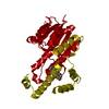
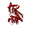
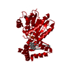
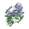
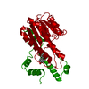
| Structure viewer | Molecule: MolmilJmol/JSmol |
|---|
- Downloads & links
-Download
| PDBx/mmCIF format | 1fdw.cif.gz | 65.9 KB | Display | PDBx/mmCIF format |
|---|---|---|---|---|
| PDB format | pdb1fdw.ent.gz | 48.5 KB | Display | PDB format |
| PDBx/mmJSON format | 1fdw.json.gz | Tree view | PDBx/mmJSON format | |
| Others |  Other downloads Other downloads |
-Validation report
| Arichive directory | https://data.pdbj.org/pub/pdb/validation_reports/fd/1fdwftp://data.pdbj.org/pub/pdb/validation_reports/fd/1fdw | HTTPS FTP |
|---|
-Related structure data
| Related structure data | 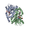 1fduC 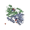 1fdvC 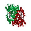 1fdtS S: Starting model for refinement C: citing same article ( |
|---|---|
| Similar structure data |

-Links
 PDBj
PDBj


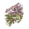
- Assembly
| Deposited unit | 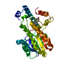
| ||||||||
|---|---|---|---|---|---|---|---|---|---|
| 1 | 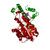
| ||||||||
| Unit cell |
|
-Components
| #1: Protein | Mass: 34963.930 Da / Num. of mol.: 1 / Mutation: H221Q Source method: isolated from a genetically manipulated source Source: (gene. exp.) Homo sapiens (human) / Cellular location: CYTOPLASM / Plasmid: PVL1393 / Cellular location (production host): CYTOPLASM / Production host:   Spodoptera frugiperda (fall armyworm) Spodoptera frugiperda (fall armyworm)References: UniProt: P14061, 17beta-estradiol 17-dehydrogenase |
|---|---|
| #2: Chemical | ChemComp-EST / 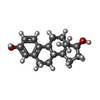  Mass: 272.382 Da / Num. of mol.: 1 / Source method: obtained synthetically / Formula: C18H24O2 / Comment: hormone*YM Mass: 272.382 Da / Num. of mol.: 1 / Source method: obtained synthetically / Formula: C18H24O2 / Comment: hormone*YM |
| #3: Water | ChemComp-HOH /  Mass: 18.015 Da / Num. of mol.: 8 / Source method: isolated from a natural source / Formula: H2O Mass: 18.015 Da / Num. of mol.: 8 / Source method: isolated from a natural source / Formula: H2O |
-Experimental details
-Experiment
| Experiment | Method: X-RAY DIFFRACTION / Number of used crystals: 1 |
|---|
- Sample preparation
| Crystal | Density Matthews: 2.46 Å3/Da / Density % sol: 50 % | |||||||||||||||||||||||||||||||||||||||||||||||||||||||||||||||||||||||||||||
|---|---|---|---|---|---|---|---|---|---|---|---|---|---|---|---|---|---|---|---|---|---|---|---|---|---|---|---|---|---|---|---|---|---|---|---|---|---|---|---|---|---|---|---|---|---|---|---|---|---|---|---|---|---|---|---|---|---|---|---|---|---|---|---|---|---|---|---|---|---|---|---|---|---|---|---|---|---|---|
| Crystal grow | pH: 7 Details: PROTEIN WAS CRYSTALLIZED FROM PEG 4000 30%, 100 MM HEPES BUFFER PH 7.0, 100 MM MGCL2, 0.5 MM ESTRADIOL, PROPANE DIOL 2-4% | |||||||||||||||||||||||||||||||||||||||||||||||||||||||||||||||||||||||||||||
| Crystal grow | *PLUS pH: 7.5 / Method: unknown | |||||||||||||||||||||||||||||||||||||||||||||||||||||||||||||||||||||||||||||
| Components of the solutions | *PLUS
|
-Data collection
| Diffraction | Mean temperature: 300 K |
|---|---|
| Diffraction source | Source: ROTATING ANODE / Type: RIGAKU RUH2R / Wavelength: 1.5418 |
| Detector | Type: SIEMENS-NICOLET X100 / Detector: AREA DETECTOR / Date: Apr 1, 1994 / Details: COLLIMATOR |
| Radiation | Monochromator: GRAPHITE(002) / Monochromatic (M) / Laue (L): M / Scattering type: x-ray |
| Radiation wavelength | Wavelength: 1.5418 Å / Relative weight: 1 |
| Reflection | Resolution: 2.7→35.8 Å / Num. obs: 6352 / % possible obs: 65.2 % / Observed criterion σ(I): 0 / Redundancy: 2.54 % / Rsym value: 0.071 |
| Reflection shell | Resolution: 2.7→2.83 Å |
| Reflection | *PLUS Rmerge(I) obs: 0.071 |
- Processing
| Software |
| ||||||||||||||||||||||||||||||||||||||||||||||||||||||||||||||||||||||||||||||||||||
|---|---|---|---|---|---|---|---|---|---|---|---|---|---|---|---|---|---|---|---|---|---|---|---|---|---|---|---|---|---|---|---|---|---|---|---|---|---|---|---|---|---|---|---|---|---|---|---|---|---|---|---|---|---|---|---|---|---|---|---|---|---|---|---|---|---|---|---|---|---|---|---|---|---|---|---|---|---|---|---|---|---|---|---|---|---|
| Refinement | Method to determine structure: MOLECULAR REPLACEMENT Starting model: PDB ENTRY 1FDT Resolution: 2.7→10 Å / σ(F): 2 Details: THE LOOP 191 - 197 HAS BEEN EXCLUDED FROM THE COORDINATE FILE.
| ||||||||||||||||||||||||||||||||||||||||||||||||||||||||||||||||||||||||||||||||||||
| Displacement parameters | Biso mean: 16.29 Å2 | ||||||||||||||||||||||||||||||||||||||||||||||||||||||||||||||||||||||||||||||||||||
| Refinement step | Cycle: LAST / Resolution: 2.7→10 Å
| ||||||||||||||||||||||||||||||||||||||||||||||||||||||||||||||||||||||||||||||||||||
| Refine LS restraints |
| ||||||||||||||||||||||||||||||||||||||||||||||||||||||||||||||||||||||||||||||||||||
| Software | *PLUS Name: REFMAC / Classification: refinement | ||||||||||||||||||||||||||||||||||||||||||||||||||||||||||||||||||||||||||||||||||||
| Refinement | *PLUS Rfactor obs: 0.178 | ||||||||||||||||||||||||||||||||||||||||||||||||||||||||||||||||||||||||||||||||||||
| Solvent computation | *PLUS | ||||||||||||||||||||||||||||||||||||||||||||||||||||||||||||||||||||||||||||||||||||
| Displacement parameters | *PLUS |
