 Movie
Movie Controller

[English] 日本語
 Yorodumi
- PDB-3gh8: Crystal structure of Mus musculus iodotyrosine deiodinase (IYD) b... -
+ Open data

- Basic information
| Entry | Database: PDB / ID: 3gh8 | ||||||
|---|---|---|---|---|---|---|---|
| Title | Crystal structure of Mus musculus iodotyrosine deiodinase (IYD) bound to FMN and di-iodotyrosine (DIT) | ||||||
 Components Components | Iodotyrosine dehalogenase 1 | ||||||
 Keywords Keywords | OXIDOREDUCTASE / IYD / iodide salvage / flavoprotein / di-iodotyrosine / DIT / FMN / Membrane / NADP / Transmembrane | ||||||
| Function / homology |  Function and homology information Function and homology informationThyroxine biosynthesis / iodotyrosine deiodinase / iodotyrosine deiodinase activity / : / thyroid hormone metabolic process / cytoplasmic vesicle membrane / FMN binding / oxidoreductase activity / nucleoplasm / plasma membrane Similarity search - Function | ||||||
| Biological species |  | ||||||
| Method |  X-RAY DIFFRACTION / SYNCHROTRON / MOLECULAR REPLACEMENT / Resolution: 2.61 Å X-RAY DIFFRACTION / SYNCHROTRON / MOLECULAR REPLACEMENT / Resolution: 2.61 Å | ||||||
 Authors Authors | Thomas, S.R. / McTamney, P.M. / Adler, J.M. / LaRonde-LeBlanc, N. / Rokita, S.E. | ||||||
 Citation Citation | Journal: J.Biol.Chem. / Year: 2009 Title: Crystal structure of iodotyrosine deiodinase, a novel flavoprotein responsible for iodide salvage in thyroid glands. Authors: Thomas, S.R. / McTamney, P.M. / Adler, J.M. / Laronde-Leblanc, N. / Rokita, S.E. | ||||||
| History |
|
- Structure visualization
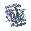
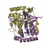
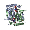
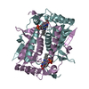
| Structure viewer | Molecule: MolmilJmol/JSmol |
|---|
- Downloads & links
-Download
| PDBx/mmCIF format | 3gh8.cif.gz | 374.3 KB | Display | PDBx/mmCIF format |
|---|---|---|---|---|
| PDB format | pdb3gh8.ent.gz | 305.2 KB | Display | PDB format |
| PDBx/mmJSON format | 3gh8.json.gz | Tree view | PDBx/mmJSON format | |
| Others |  Other downloads Other downloads |
-Validation report
| Arichive directory | https://data.pdbj.org/pub/pdb/validation_reports/gh/3gh8ftp://data.pdbj.org/pub/pdb/validation_reports/gh/3gh8 | HTTPS FTP |
|---|
-Related structure data
| Related structure data |  3gb5SC  3gfdC S: Starting model for refinement C: citing same article ( |
|---|---|
| Similar structure data |

-Links
 PDBj
PDBj- Assembly
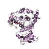
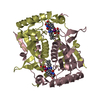
| Deposited unit | 
| ||||||||
|---|---|---|---|---|---|---|---|---|---|
| 1 | 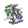
| ||||||||
| 2 | 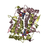
| ||||||||
| 3 | 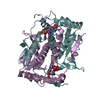
| ||||||||
| 4 | 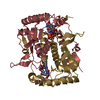
| ||||||||
| Unit cell |
|
-Components
| #1: Protein | Mass: 29987.398 Da / Num. of mol.: 8 / Fragment: UNP residues 34-285 Source method: isolated from a genetically manipulated source Source: (gene. exp.)   Spodoptera frugiperda (fall armyworm) / References: UniProt: Q9DCX8 Spodoptera frugiperda (fall armyworm) / References: UniProt: Q9DCX8#2: Chemical | ChemComp-FMN /   Mass: 456.344 Da / Num. of mol.: 8 / Source method: obtained synthetically / Formula: C17H21N4O9P Mass: 456.344 Da / Num. of mol.: 8 / Source method: obtained synthetically / Formula: C17H21N4O9P#3: Chemical | ChemComp-TYI / 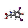  Type: L-peptide linking / Mass: 432.982 Da / Num. of mol.: 8 / Source method: obtained synthetically / Formula: C9H9I2NO3 / Comment: hormone*YM Type: L-peptide linking / Mass: 432.982 Da / Num. of mol.: 8 / Source method: obtained synthetically / Formula: C9H9I2NO3 / Comment: hormone*YM#4: Chemical |   Mass: 94.971 Da / Num. of mol.: 2 / Source method: obtained synthetically / Formula: PO4 Mass: 94.971 Da / Num. of mol.: 2 / Source method: obtained synthetically / Formula: PO4#5: Water | ChemComp-HOH / |  Mass: 18.015 Da / Num. of mol.: 300 / Source method: isolated from a natural source / Formula: H2O Mass: 18.015 Da / Num. of mol.: 300 / Source method: isolated from a natural source / Formula: H2O |
|---|
-Experimental details
-Experiment
| Experiment | Method: X-RAY DIFFRACTION / Number of used crystals: 1 |
|---|
- Sample preparation
| Crystal | Density Matthews: 2.25 Å3/Da / Density % sol: 45.26 % |
|---|---|
| Crystal grow | Temperature: 293 K / Method: vapor diffusion, hanging drop / pH: 5.5 Details: 0.2M Ammonium acetate, 45% 2-Methyl-2,4-pentanediol, 0.1M Bis-Tris pH 5.5, VAPOR DIFFUSION, HANGING DROP, temperature 293K |
-Data collection
| Diffraction | Mean temperature: 100 K |
|---|---|
| Diffraction source | Source: SYNCHROTRON / Site: APS  / Beamline: 24-ID-C / Wavelength: 0.9795 Å / Beamline: 24-ID-C / Wavelength: 0.9795 Å |
| Detector | Type: ADSC QUANTUM 315r / Detector: CCD / Date: Apr 4, 2008 |
| Radiation | Monochromator: Kohzu HLD8-24 / Protocol: SINGLE WAVELENGTH / Scattering type: x-ray |
| Radiation wavelength | Wavelength: 0.9795 Å / Relative weight: 1 |
| Reflection | Resolution: 2.6→50 Å / Num. all: 62935 / Num. obs: 62878 / % possible obs: 97.1 % / Observed criterion σ(I): 2 / Redundancy: 2.6 % / Rmerge(I) obs: 0.086 / Rsym value: 0.086 / Net I/σ(I): 9.7 |
| Reflection shell | Resolution: 2.6→2.69 Å / Redundancy: 2.5 % / Mean I/σ(I) obs: 2.4 / Num. unique all: 5984 / Rsym value: 0.295 / % possible all: 92.9 |
- Processing
| Software |
| |||||||||||||||||||||||||||||||||||||||||||||||||||||||||||||||||||||||||||||||||||||||||||||||||||||||||||||||||||||||||||||
|---|---|---|---|---|---|---|---|---|---|---|---|---|---|---|---|---|---|---|---|---|---|---|---|---|---|---|---|---|---|---|---|---|---|---|---|---|---|---|---|---|---|---|---|---|---|---|---|---|---|---|---|---|---|---|---|---|---|---|---|---|---|---|---|---|---|---|---|---|---|---|---|---|---|---|---|---|---|---|---|---|---|---|---|---|---|---|---|---|---|---|---|---|---|---|---|---|---|---|---|---|---|---|---|---|---|---|---|---|---|---|---|---|---|---|---|---|---|---|---|---|---|---|---|---|---|---|
| Refinement | Method to determine structure: MOLECULAR REPLACEMENT Starting model: PDB entry 3GB5 Resolution: 2.61→30 Å / Cor.coef. Fo:Fc: 0.951 / Cor.coef. Fo:Fc free: 0.893 / Occupancy max: 1 / Occupancy min: 0.5 / SU B: 15.028 / SU ML: 0.266 / TLS residual ADP flag: LIKELY RESIDUAL / Cross valid method: THROUGHOUT / σ(F): 0 / ESU R: 9.88 / ESU R Free: 0.354 / Stereochemistry target values: MAXIMUM LIKELIHOOD Details: HYDROGENS HAVE BEEN ADDED IN THE RIDING POSITIONS. The chirality errors and angles outside Ramachandran plot are due to poor density in flexible regions.
| |||||||||||||||||||||||||||||||||||||||||||||||||||||||||||||||||||||||||||||||||||||||||||||||||||||||||||||||||||||||||||||
| Solvent computation | Ion probe radii: 0.8 Å / Shrinkage radii: 0.8 Å / VDW probe radii: 1.2 Å / Solvent model: MASK | |||||||||||||||||||||||||||||||||||||||||||||||||||||||||||||||||||||||||||||||||||||||||||||||||||||||||||||||||||||||||||||
| Displacement parameters | Biso max: 112.41 Å2 / Biso mean: 35.532 Å2 / Biso min: 8.36 Å2
| |||||||||||||||||||||||||||||||||||||||||||||||||||||||||||||||||||||||||||||||||||||||||||||||||||||||||||||||||||||||||||||
| Refinement step | Cycle: LAST / Resolution: 2.61→30 Å
| |||||||||||||||||||||||||||||||||||||||||||||||||||||||||||||||||||||||||||||||||||||||||||||||||||||||||||||||||||||||||||||
| Refine LS restraints |
| |||||||||||||||||||||||||||||||||||||||||||||||||||||||||||||||||||||||||||||||||||||||||||||||||||||||||||||||||||||||||||||
| LS refinement shell | Resolution: 2.61→2.67 Å / Total num. of bins used: 20
| |||||||||||||||||||||||||||||||||||||||||||||||||||||||||||||||||||||||||||||||||||||||||||||||||||||||||||||||||||||||||||||
| Refinement TLS params. | Method: refined / Refine-ID: X-RAY DIFFRACTION
| |||||||||||||||||||||||||||||||||||||||||||||||||||||||||||||||||||||||||||||||||||||||||||||||||||||||||||||||||||||||||||||
| Refinement TLS group |
|
