 Movie
Movie Controller

[English] 日本語
 Yorodumi
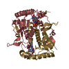
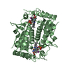
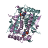
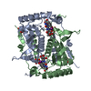
- PDB-5yak: The crystal structure of human IYD Thr239 mutant with ligand 3-Fl... -
+ Open data

- Basic information
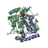
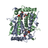
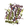
| Entry | Database: PDB / ID: 5yak | ||||||
|---|---|---|---|---|---|---|---|
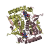
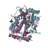
| Title | The crystal structure of human IYD Thr239 mutant with ligand 3-Fluorotyrosine (F-Tyr) | ||||||
 Components Components | Iodotyrosine deiodinase 1 | ||||||
 Keywords Keywords | OXIDOREDUCTASE / hIYD / FMN / MFT / T239A mutant | ||||||
| Function / homology |  Function and homology information Function and homology informationiodotyrosine deiodinase / iodotyrosine deiodinase activity / Thyroxine biosynthesis / : / thyroid hormone metabolic process / cytoplasmic vesicle membrane / FMN binding / oxidoreductase activity / plasma membrane Similarity search - Function | ||||||
| Biological species |  Homo sapiens (human) Homo sapiens (human) | ||||||
| Method |  X-RAY DIFFRACTION / SYNCHROTRON / MOLECULAR REPLACEMENT / Resolution: 2.3 Å X-RAY DIFFRACTION / SYNCHROTRON / MOLECULAR REPLACEMENT / Resolution: 2.3 Å | ||||||
 Authors Authors | Hu, J.M. / Rokita, S.E. / Schlessman, J. | ||||||
| Funding support |  United States, 1items United States, 1items
| ||||||
 Citation Citation | Journal: Protein Sci. / Year: 2019 Title: Redox control of iodotyrosine deiodinase Authors: Hu, J. / Su, Q. / Schlessman, J.L. / Rokita, S.E. #1: Journal: To Be PublishedTitle: The role of Thr239 on the catalysis of hIYD Authors: Hu, J.M. / Rokita, S.E. | ||||||
| History |
|
- Structure visualization
| Structure viewer | Molecule: MolmilJmol/JSmol |
|---|
- Downloads & links
-Download
| PDBx/mmCIF format | 5yak.cif.gz | 286 KB | Display | PDBx/mmCIF format |
|---|---|---|---|---|
| PDB format | pdb5yak.ent.gz | 229.5 KB | Display | PDB format |
| PDBx/mmJSON format | 5yak.json.gz | Tree view | PDBx/mmJSON format | |
| Others |  Other downloads Other downloads |
-Validation report
| Arichive directory | https://data.pdbj.org/pub/pdb/validation_reports/ya/5yakftp://data.pdbj.org/pub/pdb/validation_reports/ya/5yak | HTTPS FTP |
|---|
-Related structure data
| Related structure data | 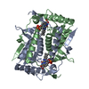 4ttcS S: Starting model for refinement |
|---|---|
| Similar structure data |

-Links
 PDBj
PDBj- Assembly
| Deposited unit | 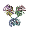
| ||||||||
|---|---|---|---|---|---|---|---|---|---|
| 1 | 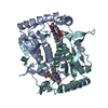
| ||||||||
| 2 | 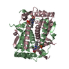
| ||||||||
| 3 | 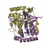
| ||||||||
| Unit cell |
|
-Components
| #1: Protein | Mass: 30537.951 Da / Num. of mol.: 6 / Fragment: UNP residues 32-289 / Mutation: T239A Source method: isolated from a genetically manipulated source Details: FMN(flavin mononucleotide) YOF(3-monofluorotyrosine) Source: (gene. exp.) Homo sapiens (human) / Gene: IYD, C6orf71, DEHAL1 / Plasmid: pET28-SUMO / Details (production host): sumo fusion N-terminal / Production host:  #2: Chemical | ChemComp-FMN /   Mass: 456.344 Da / Num. of mol.: 6 / Source method: isolated from a natural source / Formula: C17H21N4O9P / Feature type: SUBJECT OF INVESTIGATION Mass: 456.344 Da / Num. of mol.: 6 / Source method: isolated from a natural source / Formula: C17H21N4O9P / Feature type: SUBJECT OF INVESTIGATION#3: Chemical | ChemComp-YOF / 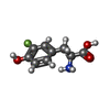  Type: L-peptide linking / Mass: 199.179 Da / Num. of mol.: 6 / Source method: obtained synthetically / Formula: C9H10FNO3 Type: L-peptide linking / Mass: 199.179 Da / Num. of mol.: 6 / Source method: obtained synthetically / Formula: C9H10FNO3#4: Water | ChemComp-HOH / |  Mass: 18.015 Da / Num. of mol.: 297 / Source method: isolated from a natural source / Formula: H2O Mass: 18.015 Da / Num. of mol.: 297 / Source method: isolated from a natural source / Formula: H2O |
|---|
-Experimental details
-Experiment
| Experiment | Method: X-RAY DIFFRACTION / Number of used crystals: 1 |
|---|
- Sample preparation
| Crystal | Density Matthews: 2.61 Å3/Da / Density % sol: 52.89 % |
|---|---|
| Crystal grow | Temperature: 293.15 K / Method: vapor diffusion, hanging drop Details: 0.17 M sodium acetate, 85 mM Tris-HCl (pH 8.5), 22.5 % w/v polyethylene glycol 4,000 and 15 % glycerol. PH range: 7.4-8.5 |
-Data collection
| Diffraction | Mean temperature: 100 K |
|---|---|
| Diffraction source | Source: SYNCHROTRON / Site: NSLS / Beamline: X25 / Wavelength: 1.1 Å |
| Detector | Type: ADSC QUANTUM 315 / Detector: CCD / Date: Feb 18, 2014 |
| Radiation | Protocol: SINGLE WAVELENGTH / Monochromatic (M) / Laue (L): M / Scattering type: x-ray |
| Radiation wavelength | Wavelength: 1.1 Å / Relative weight: 1 |
| Reflection | Resolution: 2.3→46.52 Å / Num. obs: 82734 / % possible obs: 100 % / Redundancy: 8 % / Net I/σ(I): 41.5 |
| Reflection shell | Resolution: 2.3→2.36 Å / Redundancy: 8 % / Rmerge(I) obs: 0.093 / Rsym value: 0.084 / % possible all: 100 |
- Processing
| Software |
| ||||||||||||||||||||||||||||||||||||||||||||||||||||||||||||||||||||||||||||||||||||||||||||||||||||||||||||||||||||||||||||||||||||||||||||||||||||||||||||||||||||||||||||||||||||||
|---|---|---|---|---|---|---|---|---|---|---|---|---|---|---|---|---|---|---|---|---|---|---|---|---|---|---|---|---|---|---|---|---|---|---|---|---|---|---|---|---|---|---|---|---|---|---|---|---|---|---|---|---|---|---|---|---|---|---|---|---|---|---|---|---|---|---|---|---|---|---|---|---|---|---|---|---|---|---|---|---|---|---|---|---|---|---|---|---|---|---|---|---|---|---|---|---|---|---|---|---|---|---|---|---|---|---|---|---|---|---|---|---|---|---|---|---|---|---|---|---|---|---|---|---|---|---|---|---|---|---|---|---|---|---|---|---|---|---|---|---|---|---|---|---|---|---|---|---|---|---|---|---|---|---|---|---|---|---|---|---|---|---|---|---|---|---|---|---|---|---|---|---|---|---|---|---|---|---|---|---|---|---|---|
| Refinement | Method to determine structure: MOLECULAR REPLACEMENT Starting model: 4TTC Resolution: 2.3→46.52 Å / Cor.coef. Fo:Fc: 0.96 / Cor.coef. Fo:Fc free: 0.939 / Cross valid method: THROUGHOUT / ESU R: 0.244 / ESU R Free: 0.199 / Details: HYDROGENS HAVE BEEN ADDED IN THE RIDING POSITIONS
| ||||||||||||||||||||||||||||||||||||||||||||||||||||||||||||||||||||||||||||||||||||||||||||||||||||||||||||||||||||||||||||||||||||||||||||||||||||||||||||||||||||||||||||||||||||||
| Solvent computation | Ion probe radii: 0.8 Å / Shrinkage radii: 0.8 Å / VDW probe radii: 1.2 Å | ||||||||||||||||||||||||||||||||||||||||||||||||||||||||||||||||||||||||||||||||||||||||||||||||||||||||||||||||||||||||||||||||||||||||||||||||||||||||||||||||||||||||||||||||||||||
| Displacement parameters | Biso mean: 48.406 Å2
| ||||||||||||||||||||||||||||||||||||||||||||||||||||||||||||||||||||||||||||||||||||||||||||||||||||||||||||||||||||||||||||||||||||||||||||||||||||||||||||||||||||||||||||||||||||||
| Refinement step | Cycle: 1 / Resolution: 2.3→46.52 Å
| ||||||||||||||||||||||||||||||||||||||||||||||||||||||||||||||||||||||||||||||||||||||||||||||||||||||||||||||||||||||||||||||||||||||||||||||||||||||||||||||||||||||||||||||||||||||
| Refine LS restraints |
|