 Movie
Movie Controller


+ Open data

- Basic information
| Entry |  Database: PDB chemical components / ID: YOF Database: PDB chemical components / ID: YOF |
|---|---|
| Name | Name: |
-Chemical information
| Composition |  | ||||||
|---|---|---|---|---|---|---|---|
| Others | Type: L-PEPTIDE LINKING / PDB classification: HETAIN / One letter code: Y / Three letter code: YOF / Model coordinates PDB-ID: 3FYG / Parent comp.: TYR | ||||||
| History |
| ||||||
 External links External links | UniChem / ChemSpider / Brenda / ChEBI / ChEMBL / CompTox / DrugBank / PubChem / SureChEMBL / Wikipedia search / Google search |

- Structure visualization
| Structure viewer | Molecule:  MolmilJmol/JSmol MolmilJmol/JSmol |
|---|
-Details
-SMILES
| ACDLabs 10.04 | | CACTVS 3.341 | OpenEye OEToolkits 1.5.0 | |
|---|
-SMILES CANONICAL
| CACTVS 3.341 | | OpenEye OEToolkits 1.5.0 | |
|---|
-InChI
| InChI 1.03 |
|---|
-InChIKey
| InChI 1.03 |
|---|
-SYSTEMATIC NAME
| ACDLabs 10.04 | | OpenEye OEToolkits 1.5.0 | ( | |
|---|
-PDB entries
Showing all 6 items
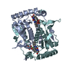
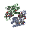
PDB-5yak: 
The crystal structure of human IYD Thr239 mutant with ligand 3-Fluorotyrosine (F-Tyr)

PDB-6mpd:
Citrobacter freundii tyrosine phenol-lyase complexed with 4-hydroxypyridine and aminoacrylate from 3-F-L-tyrosine
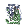
PDB-6q1b:
Crystal structure of oxidized iodotyrosine deiodinase (IYD) bound to FMN and 3-fluoro-L-tyrosine
PDB-7kqs:
A 1.68-A resolution 3-fluoro-L-tyrosine bound crystal structure of heme-dependent tyrosine hydroxylase

PDB-7kqt:
A 1.84-A resolution crystal structure of heme-dependent L-tyrosine hydroxylase in complex with 3-fluoro-L-tyrosine and cyanide

PDB-7kqu:
A 1.58-A resolution crystal structure of ferric-hydroperoxo intermediate of L-tyrosine hydroxylase in complex with 3-fluoro-L-tyrosine
