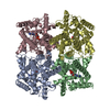
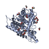
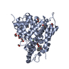
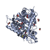
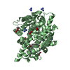
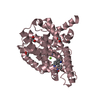
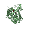
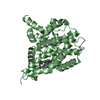
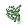
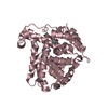
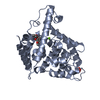
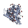
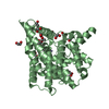
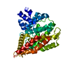
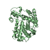
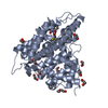
[English] 日本語
Yorodumi
- PDB-3sl8: Crystal structure of the catalytic domain of PDE4D2 with compound 10o -

+
Open data


ID or keywords:

Loading...

-
Basic information

Entry
Database: PDB / ID: 3sl8
TitleCrystal structure of the catalytic domain of PDE4D2 with compound 10o
ComponentscAMP-specific 3',5'-cyclic phosphodiesterase 4D
KeywordsHydrolase/Hydrolase Inhibitor / catalytic mechanism / cAMP hydrolysis / Hydrolase-Hydrolase Inhibitor complex
Function / homology
Function and homology information


signaling receptor regulator activity / negative regulation of relaxation of cardiac muscle / negative regulation of heart contraction / 3',5'-cyclic-AMP phosphodiesterase / positive regulation of interleukin-5 production / regulation of cardiac muscle cell contraction / establishment of endothelial barrier / negative regulation of cAMP-mediated signaling / beta-2 adrenergic receptor binding / regulation of calcium ion transmembrane transport via high voltage-gated calcium channel ...signaling receptor regulator activity / negative regulation of relaxation of cardiac muscle / negative regulation of heart contraction / 3',5'-cyclic-AMP phosphodiesterase / positive regulation of interleukin-5 production / regulation of cardiac muscle cell contraction / establishment of endothelial barrier / negative regulation of cAMP-mediated signaling / beta-2 adrenergic receptor binding / regulation of calcium ion transmembrane transport via high voltage-gated calcium channel / positive regulation of heart rate / heterocyclic compound binding / adrenergic receptor signaling pathway / voltage-gated calcium channel complex / regulation of cell communication by electrical coupling involved in cardiac conduction / cAMP catabolic process / calcium channel regulator activity / cAMP-mediated signaling / 3',5'-cyclic-nucleotide phosphodiesterase activity / 3',5'-cyclic-AMP phosphodiesterase activity / DARPP-32 events / negative regulation of peptidyl-serine phosphorylation / regulation of release of sequestered calcium ion into cytosol by sarcoplasmic reticulum / cAMP binding / cellular response to cAMP / cellular response to epinephrine stimulus / calcium channel complex / positive regulation of interleukin-2 production / regulation of heart rate / positive regulation of type II interferon production / ATPase binding / T cell receptor signaling pathway / G alpha (s) signalling events / scaffold protein binding / transmembrane transporter binding / apical plasma membrane / centrosome / perinuclear region of cytoplasm / enzyme binding / signal transduction / membrane / metal ion binding / nucleus / plasma membrane / cytosol
Similarity search - Function
Phosphodiesterase 4 upstream conserved regions (UCR) / Phosphodiesterase 4 upstream conserved regions (UCR) / Catalytic domain of cyclic nucleotide phosphodiesterase 4b2b / 3'5'-cyclic nucleotide phosphodiesterase, catalytic domain / 3'5'-cyclic nucleotide phosphodiesterase / 3'5'-cyclic nucleotide phosphodiesterase, catalytic domain / 3'5'-cyclic nucleotide phosphodiesterase, conserved site / 3'5'-cyclic nucleotide phosphodiesterase, catalytic domain superfamily / 3'5'-cyclic nucleotide phosphodiesterase / 3'5'-cyclic nucleotide phosphodiesterase domain signature. ...Phosphodiesterase 4 upstream conserved regions (UCR) / Phosphodiesterase 4 upstream conserved regions (UCR) / Catalytic domain of cyclic nucleotide phosphodiesterase 4b2b / 3'5'-cyclic nucleotide phosphodiesterase, catalytic domain / 3'5'-cyclic nucleotide phosphodiesterase / 3'5'-cyclic nucleotide phosphodiesterase, catalytic domain / 3'5'-cyclic nucleotide phosphodiesterase, conserved site / 3'5'-cyclic nucleotide phosphodiesterase, catalytic domain superfamily / 3'5'-cyclic nucleotide phosphodiesterase / 3'5'-cyclic nucleotide phosphodiesterase domain signature. / 3'5'-cyclic nucleotide phosphodiesterase domain profile. / Orthogonal Bundle / Mainly Alpha
Similarity search - Domain/homology
Chem-JN7 / DI(HYDROXYETHYL)ETHER / PHOSPHATE ION / 3',5'-cyclic-AMP phosphodiesterase 4D
Similarity search - Component
Biological speciesHomo sapiens (human)
MethodX-RAY DIFFRACTION / SYNCHROTRON / MOLECULAR REPLACEMENT / Resolution: 2.6 Å
AuthorsFeil, S.F.
CitationJournal: Bioorg.Med.Chem.Lett. / Year: 2011
Title: Thiophene inhibitors of PDE4: Crystal structures show a second binding mode at the catalytic domain of PDE4D2.
Authors: Nankervis, J.L. / Feil, S.C. / Hancock, N.C. / Zheng, Z. / Ng, H.L. / Morton, C.J. / Holien, J.K. / Ho, P.W. / Frazzetto, M.M. / Jennings, I.G. / Manallack, D.T. / Martin, T.J. / Thompson, P.E. / Parker, M.W.
History
DepositionJun 24, 2011Deposition site: RCSB / Processing site: RCSB
Revision 1.0Oct 26, 2011Provider: repository / Type: Initial release
Revision 1.1Dec 14, 2011Group: Database references
Revision 1.2Nov 8, 2017Group: Refinement description / Category: software / Item: _software.name
Revision 1.3Feb 28, 2024Group: Data collection / Database references / Derived calculations
Category: chem_comp_atom / chem_comp_bond ...chem_comp_atom / chem_comp_bond / database_2 / pdbx_struct_conn_angle / struct_conn / struct_site
Item: _database_2.pdbx_DOI / _database_2.pdbx_database_accession ..._database_2.pdbx_DOI / _database_2.pdbx_database_accession / _pdbx_struct_conn_angle.ptnr1_auth_asym_id / _pdbx_struct_conn_angle.ptnr1_auth_comp_id / _pdbx_struct_conn_angle.ptnr1_auth_seq_id / _pdbx_struct_conn_angle.ptnr1_label_asym_id / _pdbx_struct_conn_angle.ptnr1_label_atom_id / _pdbx_struct_conn_angle.ptnr1_label_comp_id / _pdbx_struct_conn_angle.ptnr1_label_seq_id / _pdbx_struct_conn_angle.ptnr2_auth_asym_id / _pdbx_struct_conn_angle.ptnr2_auth_seq_id / _pdbx_struct_conn_angle.ptnr2_label_asym_id / _pdbx_struct_conn_angle.ptnr3_auth_asym_id / _pdbx_struct_conn_angle.ptnr3_auth_comp_id / _pdbx_struct_conn_angle.ptnr3_auth_seq_id / _pdbx_struct_conn_angle.ptnr3_label_asym_id / _pdbx_struct_conn_angle.ptnr3_label_atom_id / _pdbx_struct_conn_angle.ptnr3_label_comp_id / _pdbx_struct_conn_angle.ptnr3_label_seq_id / _pdbx_struct_conn_angle.value / _struct_conn.pdbx_dist_value / _struct_conn.ptnr1_auth_asym_id / _struct_conn.ptnr1_auth_comp_id / _struct_conn.ptnr1_auth_seq_id / _struct_conn.ptnr1_label_asym_id / _struct_conn.ptnr1_label_atom_id / _struct_conn.ptnr1_label_comp_id / _struct_conn.ptnr1_label_seq_id / _struct_conn.ptnr2_auth_asym_id / _struct_conn.ptnr2_auth_comp_id / _struct_conn.ptnr2_auth_seq_id / _struct_conn.ptnr2_label_asym_id / _struct_conn.ptnr2_label_atom_id / _struct_conn.ptnr2_label_comp_id / _struct_conn.ptnr2_label_seq_id / _struct_site.pdbx_auth_asym_id / _struct_site.pdbx_auth_comp_id / _struct_site.pdbx_auth_seq_id

-
Structure visualization

Structure viewerMolecule:
MolmilJmol/JSmol

Downloads & links

-
Assembly

Deposited unit
A: cAMP-specific 3',5'-cyclic phosphodiesterase 4D
B: cAMP-specific 3',5'-cyclic phosphodiesterase 4D
C: cAMP-specific 3',5'-cyclic phosphodiesterase 4D
D: cAMP-specific 3',5'-cyclic phosphodiesterase 4D
hetero molecules


Theoretical massNumber of molelcules
Total (without water)170,88252
Polymers166,0964
Non-polymers4,78648
Water4,252236
1
A: cAMP-specific 3',5'-cyclic phosphodiesterase 4D
hetero molecules


Theoretical massNumber of molelcules
Total (without water)43,02818
Polymers41,5241
Non-polymers1,50417
Water181
TypeNameSymmetry operationNumber
identity operation1_555x,y,z1
MethodPISA
2
B: cAMP-specific 3',5'-cyclic phosphodiesterase 4D
hetero molecules


Theoretical massNumber of molelcules
Total (without water)42,3347
Polymers41,5241
Non-polymers8106
Water181
TypeNameSymmetry operationNumber
identity operation1_555x,y,z1
MethodPISA
3
C: cAMP-specific 3',5'-cyclic phosphodiesterase 4D
hetero molecules


Theoretical massNumber of molelcules
Total (without water)42,4759
Polymers41,5241
Non-polymers9518
Water181
TypeNameSymmetry operationNumber
identity operation1_555x,y,z1
MethodPISA
4
D: cAMP-specific 3',5'-cyclic phosphodiesterase 4D
hetero molecules


Theoretical massNumber of molelcules
Total (without water)43,04418
Polymers41,5241
Non-polymers1,52017
Water181
TypeNameSymmetry operationNumber
identity operation1_555x,y,z1
MethodPISA
Unit cell
Length a, b, c (Å)99.140, 111.650, 161.740
Angle α, β, γ (deg.)90.000, 90.000, 90.000
Int Tables number19
Space group name H-MP212121
DetailsAS PER THE AUTHORS THE BIOLOGICAL ASSEMBLY IS UNKNOWN

-
Components

-
Protein , 1 types, 4 molecules ABCD

#1: Protein
cAMP-specific 3',5'-cyclic phosphodiesterase 4D / DPDE3 / PDE43


Mass: 41523.887 Da / Num. of mol.: 4 / Fragment: Catalytic domain, UNP residues 381-741
Source method: isolated from a genetically manipulated source
Source: (gene. exp.) Homo sapiens (human) / Gene: PDE4D, DPDE3 / Plasmid: pET15b / Production host: Escherichia coli (E. coli) / Strain (production host): BL21(DE3) Star Rosetta
References: UniProt: Q08499, 3',5'-cyclic-nucleotide phosphodiesterase

-
Non-polymers , 7 types, 284 molecules

#2: Chemical...
ChemComp-EDO / 1,2-ETHANEDIOL / ETHYLENE GLYCOL / Ethylene glycol


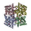
Mass: 62.068 Da / Num. of mol.: 30 / Source method: obtained synthetically / Formula: C2H6O2
#3: Chemical
ChemComp-ZN / ZINC ION


Mass: 65.409 Da / Num. of mol.: 8 / Source method: obtained synthetically / Formula: Zn
#4: Chemical ChemComp-PEG / DI(HYDROXYETHYL)ETHER / Diethylene glycol


Mass: 106.120 Da / Num. of mol.: 2 / Source method: obtained synthetically / Formula: C4H10O3
#5: Chemical
ChemComp-JN7 / 3-cyclopentyl 6-ethenyl 2-[(thiophen-2-ylacetyl)amino]-4,7-dihydrothieno[2,3-c]pyridine-3,6(5H)-dicarboxylate


Mass: 460.566 Da / Num. of mol.: 4 / Source method: obtained synthetically / Formula: C22H24N2O5S2
#6: Chemical ChemComp-PO4 / PHOSPHATE ION / Phosphate


Mass: 94.971 Da / Num. of mol.: 2 / Source method: obtained synthetically / Formula: PO4
#7: Chemical ChemComp-DMS / DIMETHYL SULFOXIDE / Dimethyl sulfoxide


Mass: 78.133 Da / Num. of mol.: 2 / Source method: obtained synthetically / Formula: C2H6OS / Comment: DMSO, precipitant*YM
#8: Water ChemComp-HOH / water / Water


Mass: 18.015 Da / Num. of mol.: 236 / Source method: isolated from a natural source / Formula: H2O

-
Experimental details

-
Experiment

ExperimentMethod: X-RAY DIFFRACTION / Number of used crystals: 1

-
Sample preparation

CrystalDensity Matthews: 2.95 Å3/Da / Density % sol: 58.24 %
Crystal growTemperature: 294 K / Method: vapor diffusion, hanging drop / pH: 7.5
Details: 20% PEG 3350, 25% (v/v) ethylene glycol, 10% (v/v) isopropanol, 100 mM HEPES, pH 7.5, vapor diffusion, hanging drop, temperature 294K

-
Data collection

DiffractionMean temperature: 100 K
Diffraction sourceSource: SYNCHROTRON / Site: Australian Synchrotron / Beamline: MX1 / Wavelength: 1 Å
DetectorType: ADSC QUANTUM 210r / Detector: CCD
RadiationMonochromator: K-B pair of biomorph mirrors / Protocol: SINGLE WAVELENGTH / Monochromatic (M) / Laue (L): M / Scattering type: x-ray
Radiation wavelengthWavelength: 1 Å / Relative weight: 1
ReflectionResolution: 2.6→20 Å / Num. obs: 53415 / % possible obs: 95.5 % / Observed criterion σ(F): 2 / Observed criterion σ(I): 2 / Redundancy: 5.7 % / Biso Wilson estimate: 46.462 Å2 / Rmerge(I) obs: 0.088 / Net I/σ(I): 16.48
Reflection shell
Resolution (Å)Rmerge(I) obsMean I/σ(I) obsNum. measured obsNum. unique obsDiffraction-ID% possible all
2.6-2.70.6663.04281775417192.2
2.7-2.80.4114.26295005015198.6
2.8-2.90.3165.33258984366198.6
2.9-30.2616.43226123806198.2
3-3.10.2137.8198613344198.4
3.1-3.20.1769.26172082897198.4
3.2-3.30.14311.44152712570197.7
3.3-3.40.12513.08134172280197.8
3.4-3.60.10515.68185963628192.7
3.6-40.07920.67262345204191.2
4-60.04531.546191010500196.3
6-100.03239.96203603517194.5
10-200.02453.254733871179.1

-
Processing

Software
NameVersionClassificationNB
XSCALEdata scaling
PHENIX1.5_2refinement
PDB_EXTRACT3.1data extraction
Blu-Icedata collection
XDSdata reduction
XDSdata scaling
MOLREPphasing
RefinementMethod to determine structure: MOLECULAR REPLACEMENT / Resolution: 2.6→19.763 Å / Occupancy max: 1 / Occupancy min: 0.29 / FOM work R set: 0.8078 / SU ML: 0.38 / σ(F): 1.99 / Phase error: 26.06 / Stereochemistry target values: ML
RfactorNum. reflection% reflectionSelection details
Rfree0.2728 2672 5 %RANDOM
Rwork0.1962 ---
obs0.2 53409 95.8 %-
all-55750 --
Solvent computationShrinkage radii: 0.9 Å / VDW probe radii: 1.11 Å / Solvent model: FLAT BULK SOLVENT MODEL / Bsol: 63.818 Å2 / ksol: 0.4 e/Å3
Displacement parametersBiso max: 118.82 Å2 / Biso mean: 43.0122 Å2 / Biso min: 12.39 Å2
Baniso -1Baniso -2Baniso -3
1-1.6253 Å2-0 Å20 Å2
2---4.2818 Å2-0 Å2
3---2.6566 Å2
Refinement stepCycle: LAST / Resolution: 2.6→19.763 Å
ProteinNucleic acidLigandSolventTotal
Num. atoms10514 0 284 236 11034
Refine LS restraints
Refine-IDTypeDev idealNumber
X-RAY DIFFRACTIONf_bond_d0.00810994
X-RAY DIFFRACTIONf_angle_d1.16614871
X-RAY DIFFRACTIONf_chiral_restr0.0771693
X-RAY DIFFRACTIONf_plane_restr0.0071898
X-RAY DIFFRACTIONf_dihedral_angle_d20.4784033
LS refinement shell

Refine-ID: X-RAY DIFFRACTION / Total num. of bins used: 19

Resolution (Å)Rfactor RfreeNum. reflection RfreeRfactor RworkNum. reflection RworkNum. reflection all% reflection obs (%)
2.6-2.64720.26521410.19072667280898
2.6472-2.6980.41411250.30592367249286
2.698-2.75290.35091450.24242739288499
2.7529-2.81260.2971410.20212705284699
2.8126-2.87780.26151440.18452723286798
2.8778-2.94960.26781420.19322700284298
2.9496-3.0290.31111430.1912720286398
3.029-3.11780.30941450.20292748289398
3.1178-3.21810.27291420.18422704284698
3.2181-3.33260.27111430.18942707285098
3.3326-3.46530.33141330.21732546267992
3.4653-3.62210.26391400.20672672281296
3.6221-3.81180.31331370.22122592272993
3.8118-4.04870.28331320.21742503263590
4.0487-4.35820.24191430.16772713285697
4.3582-4.79110.2411420.15712710285296
4.7911-5.47150.22011440.1722723286796
5.4715-6.84560.26581420.19222712285495
6.8456-19.7630.19821480.15452786293494

+
About Yorodumi

-
News

-
Feb 9, 2022. New format data for meta-information of EMDB entries

New format data for meta-information of EMDB entries

  • Version 3 of the EMDB header file is now the official format.
  • The previous official version 1.9 will be removed from the archive.

Related info.:EMDB header

External links:wwPDB to switch to version 3 of the EMDB data model

-
Aug 12, 2020. Covid-19 info

Covid-19 info

URL: https://pdbj.org/emnavi/covid19.php

New page: Covid-19 featured information page in EM Navigator.

Related info.:Covid-19 info / Mar 5, 2020. Novel coronavirus structure data

+
Mar 5, 2020. Novel coronavirus structure data

Novel coronavirus structure data

Related info.:Yorodumi Speices / Aug 12, 2020. Covid-19 info

External links:COVID-19 featured content - PDBj / Molecule of the Month (242):Coronavirus Proteases

+
Jan 31, 2019. EMDB accession codes are about to change! (news from PDBe EMDB page)

EMDB accession codes are about to change! (news from PDBe EMDB page)

  • The allocation of 4 digits for EMDB accession codes will soon come to an end. Whilst these codes will remain in use, new EMDB accession codes will include an additional digit and will expand incrementally as the available range of codes is exhausted. The current 4-digit format prefixed with “EMD-” (i.e. EMD-XXXX) will advance to a 5-digit format (i.e. EMD-XXXXX), and so on. It is currently estimated that the 4-digit codes will be depleted around Spring 2019, at which point the 5-digit format will come into force.
  • The EM Navigator/Yorodumi systems omit the EMD- prefix.

Related info.:Q: What is EMD? / ID/Accession-code notation in Yorodumi/EM Navigator

External links:EMDB Accession Codes are Changing Soon! / Contact to PDBj

+
Jul 12, 2017. Major update of PDB

Major update of PDB

  • wwPDB released updated PDB data conforming to the new PDBx/mmCIF dictionary.
  • This is a major update changing the version number from 4 to 5, and with Remediation, in which all the entries are updated.
  • In this update, many items about electron microscopy experimental information are reorganized (e.g. em_software).
  • Now, EM Navigator and Yorodumi are based on the updated data.

External links:wwPDB Remediation / Enriched Model Files Conforming to OneDep Data Standards Now Available in the PDB FTP Archive

-
Yorodumi

Thousand views of thousand structures

  • Yorodumi is a browser for structure data from EMDB, PDB, SASBDB, etc.
  • This page is also the successor to EM Navigator detail page, and also detail information page/front-end page for Omokage search.
  • The word "yorodu" (or yorozu) is an old Japanese word meaning "ten thousand". "mi" (miru) is to see.

Related info.:EMDB / PDB / SASBDB / Comparison of 3 databanks / Yorodumi Search / Aug 31, 2016. New EM Navigator & Yorodumi / Yorodumi Papers / Jmol/JSmol / Function and homology information / Changes in new EM Navigator and Yorodumi

Read more